Image Gallery: Normal vs Abnormal Ear Cytology
Elizabeth R. May, DVM, DACVD, University of Tennessee
Ear cytology provides an abundance of useful clinical information that can easily be obtained with in-house testing. When combined with otoscopic examination findings, cytology is an efficient tool in helping clinicians make a diagnosis and assess treatment response (ie, whether to discontinue or change therapy). Cytology samples should be examined under low power magnification (100×) to identify cell types and high power magnification (1000×) to identify infectious organisms, especially bacteria.

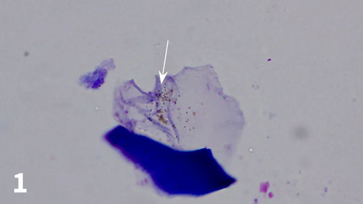
FIGURE 1
Keratinocyte with melanin granules (modified Wright’s stain; original magnification, 1000×). Melanin granules (white arrow) are frequently mistaken for rod-shaped bacteria. Note that the granules are not in chains or clumps but are organized in a haphazard fashion. Melanin granules are often refractile when examined using the fine focus knob of the microscope; note that the granules are light greenish-brown, whereas bacteria would take on a purple color when stained.
Related Articles: Chronic Otitis Externa, Flushing the External Ear Canal, Pseudomonas Otitis Infection in Cats and Dogs
Editor's note: This article was originally published in June 2016 as "Image Gallery: Ear Cytology"