Image Gallery: Dental Radiography
Jan Bellows, DVM, FAVD, DAVDC, DABVP, All Pets Dental, Weston, Florida
Along with probing and tooth-by-tooth clinical examination under anesthesia, dental radiography is an essential step in providing the best oral care for veterinary patients. This image gallery reviews common dental radiographic findings, including periapical lucency, internal and external tooth resorption, juvenile dental abnormalities, and pulpal calcification.
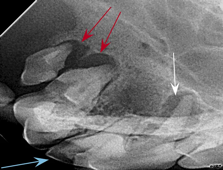
FIGURE 1: Periapical lucency
Periapical lucency develops secondary to bone loss around the tooth root caused by endodontic, neoplastic, or periodontal disease. In this dog, periapical lucency of the maxillary first and second molars (red arrows) developed secondary to periodontal disease. Periapical lucency secondary to endodontic disease (caused by exposure of the pulp from a slab fracture [blue arrow]) is also present surrounding the roots of the right maxillary fourth premolar (white arrow).
Observation of marked mobility of the molars would be expected on anesthetized examination. In this dog, the maxillary fourth premolar would likely not be mobile, as there is still bone surrounding the root apices. Extraction of the maxillary fourth premolar and first and second molars is indicated.