Image Gallery: Autopsy for the General Practitioner
Ryan N. Jennings, DVM, PhD, DACVP (Anatomic Pathology), The Ohio State University
Autopsy is a valuable diagnostic procedure that allows clinicians to identify unknown disease processes, corroborate findings of clinical or ancillary diagnostic testing, and remove tissue samples for further microscopic examination.
A sound veterinarian–owner relationship and good communication are imperative to ensure that owners understand why definitive clinical answers are not always achieved following postmortem examination.
The subject of autopsy invariably becomes relevant during emotional times for owners, including when a pet is terminally ill or recently deceased. Many owners are amenable to autopsy, particularly if it can provide definitive diagnosis (sometimes viewed as emotional closure) or help other owners who have pets with similar problems make more informed clinical decisions.
A limited, cosmetic autopsy may be the preferred choice for owners because of its less-invasive nature and the option to return the pet’s remains in a minimally altered state. The technique can be readily performed by a veterinary practitioner without expending significant financial resources. Although the nature of cosmetic autopsy limits full postmortem examination, it is an effective diagnostic procedure for determining disease pathogenesis.
The following figures pertain to the cosmetic autopsy procedure. Additional resources for further discussion of pathologic abnormalities are available.1-3
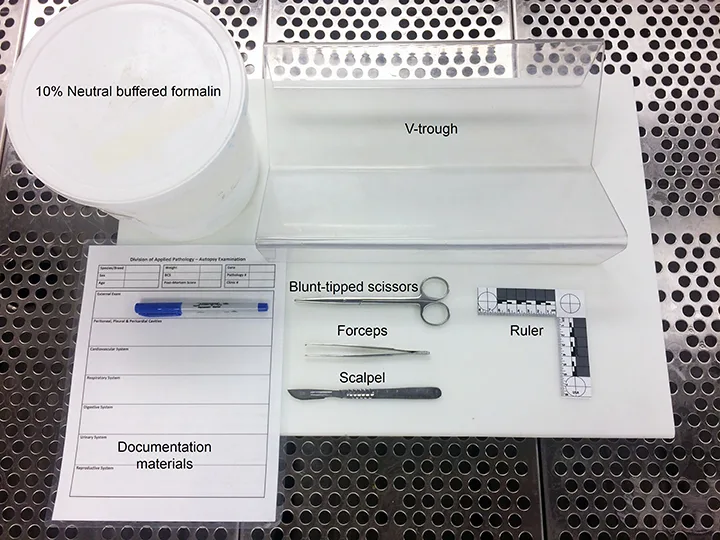
Figure 1 Materials needed for autopsy
Before beginning a postmortem examination, gather all materials needed to perform the procedure, including a container of 10% neutral buffered formalin (enough to maintain a formalin:tissue ratio of 10:1), V-trough, ruler, scalpel, forceps, blunt-tipped scissors, suture, and supplies for recording gross findings. If warranted, submit biopsied samples of formalin-fixed tissues of interest for microscopic examination at a diagnostic laboratory. Having a collection of airtight containers that withstand freezing (eg, red-top hematology tubes, bags, biopsy containers, urine specimen jars) readily available can facilitate sample collection for ancillary diagnostic testing, including toxicology, culture, and PCR testing.
Label all collected specimens with appropriate animal identification (ie, case number, animal name, client name), the attending clinician’s name, and a description of the specimen and location from which the specimen was taken (eg, fresh liver tissue).
Author's Note: The term “autopsy” is widely preferred in the pathology community, particularly to emphasize the primary goal of unifying human and veterinary medical terminology and dispelling misconceptions and misunderstandings of the procedure. Although “autopsy” is etymologically appropriate (Greek autopsia is simply to “see with one’s own eyes”), the term “necropsy” is also appropriate. Veterinary practitioners should use terminology that can be clearly communicated to and understood by clients.