Hypochloremic Metabolic Alkalosis

Teddy, a 3.5-year-old castrated male corgi, was presented for vomiting.
History. Teddy had been vomiting for 3 days. The vomit was mucoid with a green tint. His vaccinations and parasite prevention were up to date.
Examination. Teddy was quiet but alert. Poor perfusion was reflected by pale mucous membranes, prolonged capillary refill time (2 seconds), tachycardia (heart rate, 150 beats per min), and weak femoral pulse quality. He had dry mucous membranes and skin tenting. Arterial blood pressure was 90/60 mmHg; rectal temperature was 99°F. His abdomen was painful on palpation; pain was localized to the cranial right quadrant.
Laboratory Results. An emergency laboratory database was rapidly performed to identify problems that could influence immediate therapy. The Table lists the initial results.
Assessment. This patient had historical, physical, and laboratory signs consistent with hypovolemia and dehydration. There was a history of fluid loss through vomiting. Physical examination showed poor perfusion and hypotension; reduced mucous membrane moisture and skin tenting; and focal abdominal pain in the region of the proximal gastrointestinal tract, pancreas, and hepatobiliary system. Laboratory results indicated an elevated lactate level, hemoconcentration, and hypochloremic metabolic alkalosis with hypokalemia.
ASK YOURSELF ...What is the most likely cause of the patient's hypochloremic metabolic alkalosis?
A. Vomiting of gastric contentsB. Renal failureC. Toxin ingestionD. Dehydration from not drinking
What is the best crystalloid to use during volume resuscitation and rehydration of this patient?A. Balanced buffered isotonic solution, such as Normosol-R or lactated Ringer's solutionB. 3% hypertonic saline solution with potassium supplementationC. Half-strength lactated Ringer's solution with 5% dextroseD. 0.9% sodium chloride solution and potassium supplementation
CORRECT ANSWERS:A. Vomiting of gastric contentsD. 0.9% sodium chloride solution and potassium supplementation
The most common cause of hypochloremic metabolic alkalosis is vomiting of gastric fluid. Vomiting can cause a loss of hydrochloric acid (hydrogen and chloride ions), resulting in an accumulation of plasma bicarbonate (alkalemia). Because there is a reduced amount of plasma chloride (negative ion), less chloride filters from the plasma into the urine. To maintain ion equilibrium, renal tubular cells retain bicarbonate (negative ion), which exacerbates alkalemia.
In addition, hypokalemia promotes bicarbonate retention and a paradoxical aciduria. The presence of a volume-contracted state stimulates aldosterone release; this, in turn, exacerbates the existing hypokalemia and alkalosis by supporting potassium and hydrogen secretion in the tubules in exchange for sodium.
Initial Treatment. Correcting the chloride deficit with an isotonic solution containing the highest chloride level (ie, 0.9% sodium chloride: sodium, 154 mEq/L; chloride, 154 mEq/L; osmolality, 308 mOsm/L) and supplementing with potassium chloride are the most efficient ways to reduce renal retention of bicarbonate and promote intravascular volume replacement. The fluid does not need to be buffered in this situation since metabolic acidosis is not occurring.
Initial treatment requires rapid fluid bolus infusion of 0.9% sodium chloride until hypovolemia is corrected, perfusion measures are restored, and heart rate and blood pressure are normalized. The administration of a large-molecular-weight colloid, such as hetastarch (in 0.9% sodium chloride), helps to rapidly expand and retain intravascular volume and reduce the amount of crystalloid that might extravasate into third-body fluid spaces (the gastrointestinal tract in this situation). Analgesia will be necessary to remove sympathetic stimulation caused by pain.
After intravascular volume replacement, dehydration is corrected by using 0.9% sodium chloride supplemented with potassium chloride. Potassium chloride is aggressively supplemented at a rate of 0.5 to 1 mEq/kg per H until normal values are achieved. Repeated evaluation of electrolyte and pH status is necessary to prevent hyperkalemia and ensure that the therapeutic plan is appropriate. Once the hypochloremic metabolic alkalosis is corrected, an isotonic balanced crystalloid, such as Normosol-R (hospira.com) or lactated Ringer's solution may be substituted for normal saline.

Additional Diagnostics. Hypochloremic metabolic alkalosis can be caused by a pyloric outflow obstruction (foreign body, tumor, pyloric hypertrophy), gastric stasis, upper intestinal obstruction, duodenal stasis, pancreatitis, and removal of large volumes of gastric contents by nasogastric suctioning. Since the patient is a young dog with focal pain in the right upper abdominal quadrant, foreign-body obstruction and pancreatitis are the most likely causes.
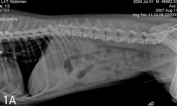
Testing should include survey radiography and abdominal ultrasonography. In this case, abdominal radiographs showed dilation of the stomach with a fluid density (Figure 1). Abdominal ultrasonography showed a very dilated and fluid-filled stomach and a proximal duodenum that exhibited little motility and contained an intraluminal foreign body (Figure 2). The foreign body had a hyperechoic surface with distal acoustic shadowing. The intestines distal to this object were normal.
Figure 1. Lateral (A) and ventrodorsal (B) abdominal radiographs showing dilation of the stomach with a fluid density and a focal gas dilation of the intestinal tract i n the right upper abdominal quadrant.
Figure 2. Abdominal ultrasonographic image of an intraluminal structure within the descending duodenum that has a hyperechoic surface with distal acoustic shadowing.
Case Management. A nasogastric tube was placed to decompress the stomach, relieve any discomfort from the distention, and prevent additional vomiting that could lead to aspiration, especially during anesthesia induction and surgery. Electrolyte and blood gas values were corrected after fluid resuscitation and rehydration. The 0.9% saline solution was discontinued, and a balanced, buffered isotonic saline solution (Normosol-R) was started to maintain intravascular volume during anesthesia and surgery. Potassium chloride was added to the crystalloids as indicated by the electrolyte panel results. Abdominal exploratory surgery uncovered a peach pit lodged at the duodenal flexure. The foreign body was removed and the patient had an uneventful recovery.
Take-Home MessageWhen pyloric outflow obstruction is suspected or hypochloremic metabolic alkalosis has been diagnosed, chloride replacement is key to correcting acid-base and electrolyte imbalance. This is efficiently accomplished by using hetastarch and 0.9% sodium chloride during fluid resuscitation and maintenance. Potassium chloride is supplemented as needed.