Horner Syndrome at a Glance
Mark Troxel, DVM, DACVIM (Neurology), Massachusetts Veterinary Referral Hospital, Woburn, Massachusetts
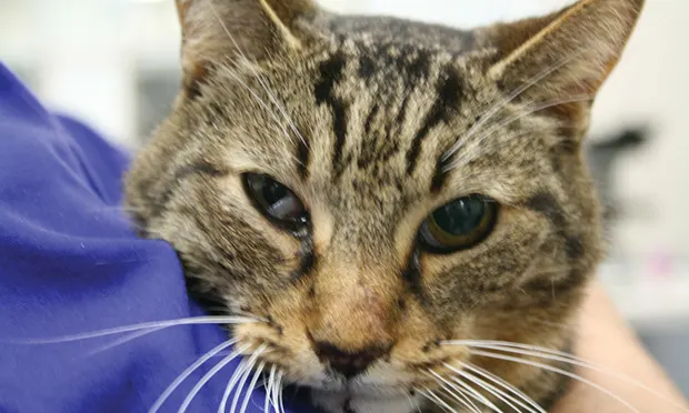
Patient showing classic signs of right-sided Horner syndrome: miosis, ptosis, enophthalmos, and elevated nictitans.
Horner syndrome (Figures 1–3) is not a disease but a myriad of clinical signs caused by sympathetic denervation to the eye. Disease affecting any portion of the sympathetic pathway can lead to ipsilateral neurologic dysfunction.
Related Article: Horner Syndrome: Identifying the Cause
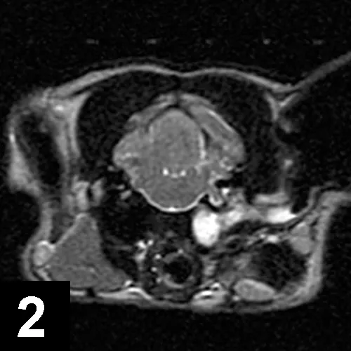
Axial T2-weighted MRI of a cat with left-sided Horner syndrome and peripheral vestibular dysfunction secondary to severe otitis.
Sympathetic innervation to the eye is a three-neuron pathway:
Upper Motor (First Order) Neuron
This cell body, located in the hypothalamus, projects axons through the brainstem and cervical spinal cord to the level of T1-T3 spinal segments.
Preganglionic (Second Order) Neurons
Axons of preganglionic neurons, which arise in the T1-T3 spinal region, leave the spinal cord through the ventral roots and pass through the cranial thorax and neck as part of the vagosympathetic trunk and synapse in the cranial cervical ganglion ventromedial to the tympanic bulla.
Related Article: Elevation of the Third Eyelid & Miosis in a Cat
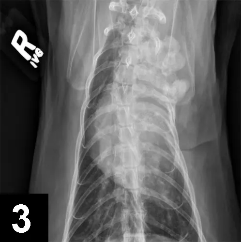
VD thoracic radiograph of a cat with left-sided Horner syndrome showing a sarcoma affecting the first 4 ribs and cranial thorax on the left, impacting preganglionic neurons.
Postganglionic (Third Order) Neurons
Postganglionic neurons originate in the cranial cervical ganglion and send axons near (ie, in dogs) or through (ie, in cats) the tympanic bulla, into the calvaria, and to the eye.