Hepatobiliary Imaging with Radiography & Ultrasonography: Liver
Lorrie Gaschen, DVM, PhD, Dr.med.vet, DECVDI, Louisiana State University
Abdominal radiography is important in the diagnosis of hepatobiliary disease, as it can help detect abnormal liver size and shape and identify mineralization or gas in the liver or intra- and extrahepatic biliary tract. Abdominal ultrasonography is the most useful noninvasive diagnostic tool for differentiating hepatic from posthepatic causes of icterus, obstructive from nonobstructive biliary disease, and diffuse from focal hepatic parenchymal disease.1
This article is part of an illustrative series on hepatobiliary imaging, including both the liver and biliary system.
The liver, gallbladder, biliary tract, duodenal papilla, and pancreas should be examined with a high-frequency transducer, and, because these structures are in the cranial abdomen, it is usually best to use a curved-array transducer with a small footprint.
Ultrasonography is useful for estimating hepatic size and evaluating internal architecture (including portal, venous, arterial, and biliary vasculature; echogenicity; and echotexture). Size, wall thickness, and contents of the gallbladder, as well as size of the bile ducts, can also be assessed. Ultrasonography is also helpful for differentiating chronic injury (eg, cirrhosis, end-stage chronic inflammatory disease) from acute liver injury based on the size, shape, and internal architecture of the liver parenchyma.2
The gallbladder wall and contents can be assessed for cholecystitis, cholelithiasis, and mucoceles via ultrasound. Ultrasound-guided tissue sampling of the liver parenchyma and cholecystocentesis is safe and often necessary due to the nonspecific nature of imaging findings.3-6

FIGURE 1A
Right lateral radiographs of a clinically normal dog (A) and an 11-year-old neutered male dachshund with diabetes mellitus (B). The dog with diabetes mellitus has a pendulous abdomen and an enlarged liver (ie, hepatomegaly) with rounded margins that extend caudal to the costal arch (B; arrows). The gastric axis (solid lines; aligned with the gastric fundus dorsally to the antrum ventrally) is parallel to the ribs in the clinically normal dog but caudally displaced in the dog with hepatomegaly. Common differential diagnoses for generalized hepatomegaly are vacuolar hepatopathy due to endocrinopathies or other metabolic disease (eg, lipidosis), infectious and noninfectious inflammatory disease, neoplasia, storage disease, and venous congestion.

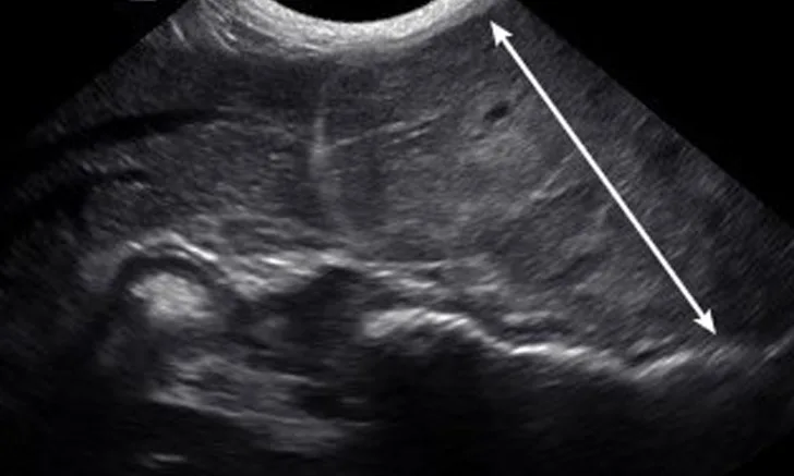
FIGURE 11A
Imaging of a 1-year-old neutered male standard poodle presented with anorexia, lethargy, vomiting, hypoalbuminemia, hypoproteinemia, and cranial abdominal organomegaly. Transverse (A) and sagittal (B) ultrasound of the liver and gallbladder (GB). The liver is enlarged (with increased distance off the stomach from the diaphragm, rounded borders, and extension ventral to the stomach), diffusely hyperechoic, and homogenous. The gallbladder is moderately distended with a normal wall and content. Radiographs demonstrate hepatomegaly and splenomegaly (C). Abdominal ultrasound images revealed splenic enlargement with a mildly mottled architecture and a solitary hypoechoic nodule (D; arrowheads). The pancreas is enlarged and lobulated with multiple hypoechoic tracts and a peripheral capsule (E; between electronic cursors [X’s]). The jejunal lymph nodes are severely enlarged (F; between electronic cursors [X’s]), more than expected for a young dog. The combination of severe hepatosplenomegaly and lymphadenomegaly make systemic disease (eg, infectious, neoplastic) most likely. The appearance of the pancreas is due to pancreatic edema secondary to hypoproteinemia. Acute liver injury was diagnosed, and lymphoma was diagnosed based on hepatic and splenic cytology.
This article is part of an illustrative series on hepatobiliary imaging. See the companion article for images of the biliary system.