Hemorrhagic Anterior Uveitis in a Labrador Retriever
Alison Clode, DVM, DACVO, Port City Veterinary Referral Hospital, Portsmouth, New Hampshire

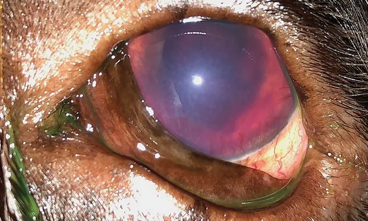
The patient’s left eye on presentation. The generalized corneal edema, superficial and deep peripheral corneal vascularization, and intraocular hemorrhage, with a visible but difficult to visualize pupil, are evident.
THE CASE
Sammy, a 9-year-old neutered male Labrador retriever, is presented for evaluation of a red, cloudy, painful left eye. The abnormal appearance to the eye was noted by the owners after Sammy had been left to play unattended with the other dog in the household for approximately 2 hours. No treatment was administered by the owners, and Sammy was presented 45 minutes after the owners first noted the abnormality.
On examination, Sammy is bright, alert, and responsive. The left eye is blepharospastic, with mild swelling of the eyelids, conjunctival hyperemia, corneal edema, hyphema, and superficial and deep peripheral corneal vascularization (Figure 1). The pupil is difficult to visualize but appears miotic as compared with the right eye. Direct pupillary light reflex (PLR) is absent, and consensual PLR in the right eye is present but subjectively decreased. Dazzle reflex (ie, reflex reaction to stimulation of the eye by a bright light) is positive, but menace response is negative in the left eye. Menace response, direct PLR, and dazzle reflex are positive in the right eye, but consensual PLR in the left eye is difficult to visualize due to hyphema in the left eye. Schirmer tear test shows >15 mm wetting/min in both eyes, fluorescein stain is negative in both eyes, and intraocular pressure (IOP), estimated by applanation tonometry, is 14 mm Hg in the right eye and 6 mm Hg in the left. The remainder of the ocular examination in the right eye, including fundic examination, is normal.
What are the next steps?
IOP = intraocular pressure, PLR = pupillary light reflex