Glass Ingestion in a Boxer: 1 Case, 2 Options
Mary Dell Deweese, DVM, Animal Medical Center, New York, New York
Karen M. Tobias, DVM, MS, DACVS, University of Tennessee

THE CASE
A 6-year-old castrated boxer is presented 24 hours after consuming a 3-lb pork roast and the glass top of a slow cooker. The owners report an episode of vomiting followed by an episode of diarrhea. The patient has a normal energy level with a mildly reduced appetite.
On digital rectal examination, there are palpable pieces of tempered glass without evidence of blood.
At presentation, the dog is bright and alert with normal vital signs. Mucous membranes are mildly hyperemic and moist with no evidence of oral ulcerations or masses. The patient is tense and nonpainful on abdominal palpation. On digital rectal examination, there are palpable pieces of tempered glass (each 1010 mm) without evidence of blood.
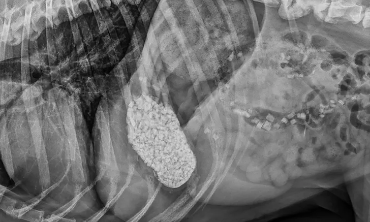
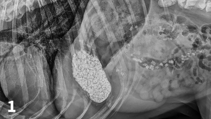
CBC reveals a leukocytosis (19.34 103/L; range, 5.10-14.00) characterized by a lymphocytosis (5.5 103/L; range, 1.4-4.6) and a mature neutrophilia (13.10 103/L; range, 2.65-9.80) with a mild thrombocytopenia (171 103/L; range, 147-243). A pancreatic-specific lipase is elevated (580 g/L; range, 0-200 g/L) and consistent with pancreatitis. Abdominal radiographs (Figures 1-3; See below) reveal a moderately distended stomach that contains a large number of irregularly shaped mineral opacities. Additional mineral opacities are present in multiple small intestinal segments as well as the colon. There is also a mild decrease in serosal detail in the mid-abdominal region.
You suspect pancreatitis and elect to admit the patient for hospitalization and supportive care.
Abdominal radiographs, repeated 14 hours later, reveal minimal aboral movement of the mineral opacities (Figures 4 and 5; see below).

Figure 1.
A right lateral abdominal radiograph taken approximately 24 hours after glass ingestion. Multiple, irregularly shaped mineral opacities are present within the stomach, small intestine, and colon.