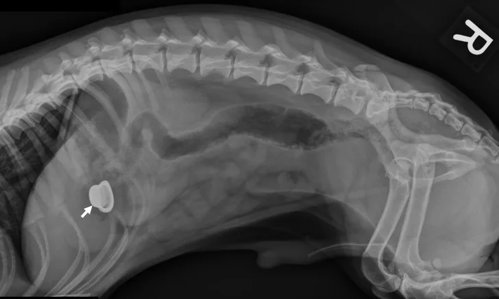
Radiographic Signs of GI Obstructions in Dogs & Cats
Eric T. Hostnik, DVM, MS, DACVR, DACVR-EDI, The Ohio State University
Michael J. Botros, DVM candidate, The Ohio State University

GI obstructions are common problems that typically require therapeutic intervention. A mechanical ileus is secondary to a physical obstruction of the intestine. Foreign bodies are among the most common causes of mechanical obstructions, the severity of which is influenced by the completeness, location, and duration of the obstruction.
Radiographs can show segmental dilation of the bowel. In dogs, intestinal diameter measurement of >1.6 times the height of the midbody of the L5 vertebra is suggestive of intestinal dilation.1 In cats, intestinal diameter measurement of >12 mm or >2 times the height of the L2 vertebra is suggestive of intestinal dilation.1 Measurements are not definitive but can act as guidelines that can be correlated with clinical signs. As the size of the intestinal dilation increases, so do the concerns for surgical obstruction.
Mild to moderate diffuse intestinal dilation (vs segmental dilation) can suggest that a functional ileus is present and can be precipitated by inflammation, metabolic derangements, or abdominal pain secondary to intestinal dilation. Small intestinal functional ileus does not typically require emergency surgery (except severe diffuse intestinal dilation caused by a mesenteric torsion).

FIGURE 1
Left lateral image showing segmental dilated gas-filled intestines (dashed arrows) with heterogeneous soft tissue in a small intestinal segment (solid arrow) in a dog. The foreign material was cloth, and the diagnosis was small intestinal mechanical obstruction. Exploratory laparotomy was performed.
Editor's note: This article originally ran in September 2021 as "GI Obstruction on Radiographs"