Gastric Dilation & GI Obstruction in Rabbits
David Eshar, DVM, DABVP (ECM), DECZM (SM & ZHM), Kansas State University
In the literature
Brezina T, Fehr M, Neumüller M, Thöle M. Acid-base-balance status and blood gas analysis in rabbits with gastric stasis and gastric dilation. J Exotic Pet Med. 2020;32:18-26.
The Research …
GI stasis is one the most common health issues in pet rabbits and often occurs secondary to an underlying medical issue.1 With the exception of physiologic ileus (eg, from a low-fiber diet), common primary problems that occur with GI stasis include gastric dilation and GI obstruction, typically of either the pylorus or duodenum. In most cases, a small ingested hair pellet is the cause of the intestinal obstruction.2 Stomach outflow obstruction can lead to caudal vena cava compression, cranial displacement of the diaphragm, activation of the sympathetic nervous system, severe pain, and reduced respiratory lung volume and heart preload. If the stomach outflow obstruction is left untreated, the pathophysiologic changes continue to worsen, leading to debilitating pain, hypovolemia, hypotension, hypothermia, disseminated intravascular coagulation, and acidosis.3-9
In addition to diagnostic imaging, clinical pathology (ie, CBC, serum chemistry profile, blood gas analysis) is important for determining a patient’s health status.3-11 For example, hyponatremia is considered a negative prognostic factor,4 and severe hyperglycemia can occur due to severe pain and stress, which can affect treatment and management decisions.5 As in other species with severe GI disturbances, blood gas analysis and acid-base status can be useful in determining appropriate treatment.
In this study of pet rabbits with gastric dilation and suspected obstruction, the authors evaluated acid-base status, electrolytes, and blood gas values, as well as how both time of presentation and therapy influenced these parameters. Prospective data from 30 rabbits were included. The resulting data suggest that acid-base balance disturbances due to gastric dilation can worsen over time without treatment. Specifically, partial pressure of carbon dioxide, partial pressure of bicarbonate, and base excess were significantly lower in rabbits presented 12 hours after the owner first noticed signs of illness as compared with rabbits presented within 6 hours. These findings strongly suggest the need for immediate veterinary care in rabbits showing reduced activity, dysphagia, and changes in fecal output.
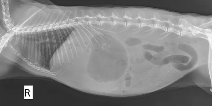
Right lateral radiograph of a 1-year-old, intact male Holland lop rabbit presented with a history of reduced activity, anorexia, and no fecal output since the previous day. Blood glucose was 328 mg/dL (reference range, 110-160 mg/dL). The stomach appears distended and fluid-filled with a gas cap, and an abnormal gas pattern can be seen in the small intestines. The combination of history, clinical signs, physical examination, hyperglycemia, and radiographic findings is highly suggestive of intestinal obstruction.
… The Takeaways
Key pearls to put into practice:
Survey radiography—including ventrodorsal, dorsoventral, and lateral views—is important in the initial screening and diagnosis of any rabbit showing GI or nonspecific clinical signs.11
Elevated blood glucose can indicate severe pain and help in the evaluation of efficacy of provided analgesics.5
Rectal temperature should always be obtained during physical examination of rabbits. Decreased rectal temperature at presentation has been shown to be a poor prognostic factor in rabbits with signs of GI dysfunction, particularly those with a temperature ≤97.9°F (36.6°C).7