Fractured or Discolored Tooth

You have asked…What is the best approach to a fractured or discolored tooth?
The expert says…
Fractured, discolored, or traumatized teeth are relatively common and can be significant sources of chronic discomfort and infection. Clinical evaluation and dental radiography can help direct treatment.
Related Article: Fractured Upper Canine Tooth
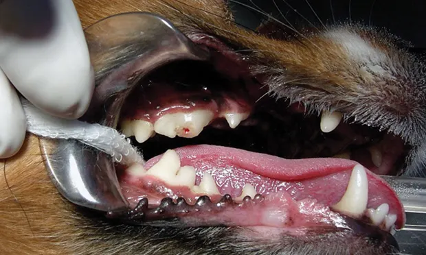
BackgroundTraumatized teeth vary from mild enamel infractions to complete avulsion. In general, tooth fractures extend through enamel then into dentin and may involve the pulp canal (Figure 1). On occasion, the tooth is not fractured, but inflammation of pulp tissue results in hemorrhage, ultimately causing pulp necrosis and tooth discoloration.
Figure 1. The right maxillary 4th premolar (ie, #108) in this patient has a complicated crown fracture (see Diagnostic Terminology).
Trauma to the tooth surface may result in enamel fractures without involving the underlying dentin, crown fractures that involve the dentin with or without pulp, crown fractures that extend along the root surface, root fractures with no crown involvement, tooth discoloration from pulpitis, or tooth luxation and/or complete avulsion. Discolored teeth are often caused by blunt trauma, with more than 90% progressing to nonvitality.1 Because pulp exposure and/or nonvital teeth are sources of chronic infection and discomfort, affected teeth need to be treated.
Related Article: Dentin Bonding: Uncomplicated Crown Fracture
Clinical Signs & EvaluationFractured teeth should be cleaned and evaluated for pulp exposures and probed for periodontal evaluation. Pulp involvement is consequential. Pulp exposure, at least initially, can be a source of acute pain. Over time, pulp tissue can become necrotic and a source of chronic infection and pain.
Related Article: To Extract or Not to Extract
Radiographic evaluation is key for diagnosis and treatment. Radiographs must be evaluated for evidence of periodontal disease, periapical rarefaction, discrepancy in pulp size (as compared with the same contralateral tooth), tooth or root resorption, and root fractures. Although it is a subjective evaluation of pulp vitality, transillumination through the crown may help detect small fractures. Vital teeth typically transilluminate as translucent, whereas nonvital teeth transilluminate with a shadow within the crown. If the contralateral tooth is considered healthy, it can be used as a reference.
Clinical Considerations & Treatment OptionsNot all fractured teeth are created equal: some may have little remaining tooth structure to preserve via root canal therapy, some may not be healthy periodontally, and others may not be mature enough for standard endodontic therapy. There are many considerations when deciding the optimum treatment. All options and potential prognoses for the affected tooth should be discussed with the client to help direct treatment decisions.
Related Article: Care of Permanent Tooth Crown Fractures in Dogs

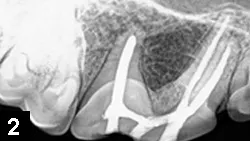
Client Expectations If clients are not interested in preserving the tooth, extraction is best; if they do wish to preserve the tooth, vital pulp therapy, standard root canal therapy (Figure 2), or surgical root canal therapy can be considered. In more complicated situations, and to include all options, consultation with a board-certified veterinary dentist may be helpful.
Figure 2. A right maxillary 4th premolar tooth (ie, #108) following standard root canal therapy (complete pulpectomy and pulp canal obturation)
Amount of Crown RemainingIf no clinical crown remains, extraction is typically ideal. Exceptions include cases in which root extraction carries a likelihood of jaw fracture or if root location (eg, areas that have undergone radiation therapy) could be problematic for extraction healing.
Tooth AgeA root must be fully formed for standard root canal therapy to have a favorable prognosis; otherwise, extraction or advanced endodontic procedures (eg, apexogenesis [if the pulp exposure is relatively recent, <1–2 weeks], apexification [see Treatment Terminology]) may be necessary.
Duration of FractureDuration of pulp exposure to oral bacteria is important; in general, the longer the pulp is exposed, the deeper and more extensive the pulp contamination. If pulp has been exposed for fewer than 48 hours or in teeth with undeveloped roots, partial coronal pulpectomy is an option; however, as standard root canal therapy has a more favorable prognosis, standard root canal therapy or extraction is usually the preferred treatment if the root is fully developed (the apex of the tooth root has closed).
Tooth HealthIf the tooth is of poor periodontal health or if the fracture extends apically along a root such that the periodontal health of the tooth is jeopardized, clients must understand the long-term prognosis in preserving the tooth is less than ideal.
Root HealthChronic pulpitis may cause resorption from the pulp tissue (ie, internal resorption). Because standard root canal therapy may stop internal resorption, this therapy is an option in some cases. Chronic inflammation in periradicular tissues can result in external resorption, making extraction ideal unless resorption is localized to the root apex. In this case, standard root canal therapy followed by surgical exposure of the involved root apex, amputation of the root tip, and retrograde placement of an appropriate filling (ie, surgical root canal therapy) is another option.
Discolored Teeth Discolored teeth usually have most of the clinical crown remaining and, if the root is otherwise healthy, are frequently excellent candidates for root canal therapy. Taking into consideration the low likelihood of a vital pulp in discolored teeth, these teeth should either be extracted or undergo standard root canal therapy.
R. MICHAEL PEAK, DVM, DAVDC, is owner of Tampa Bay Veterinary Dentistry and The Pet Dentist at Tampa Bay, referral practices in Largo and Tampa, Florida, respectively. He has served as president of the American Veterinary Dental College, chair of the examination committee, and program chair for the Veterinary Dental Forum. Dr. Peak graduated with honors from Auburn University and completed his veterinary dentistry residency at Dallas Dental Service Animal Clinic.
References
1. Localized intrinsic staining of teeth due to pulpitis and pulp necrosis in dogs. Hale FA. J Vet Dent 18:14- 20, 2001.2. Dental fracture classification. AVDC Nomenclature Committee; http://www.avdc.org/nomenclature.html#toothfracture; accessed Dec 2013.3. Principles and Practice of Endodontics, 3rd ed. Walton RE, Torabinejad M—London: Saunders Elsevier, 2002, p 390.