Forelimb Lameness in the Active Dog
Owners often bring in active dogs with complaints of forelimb lameness and commonly leave without a definitive diagnosis, departing instead with a series of nonsteroidal anti-inflammatory drugs (NSAIDs) and directions for crate rest.
Profile
Many causes of forelimb lameness in the dog need to be thoroughly examined. These conditions include bicipital tenosynovitis, supraspinatus tendinopathy, medial shoulder instability, and collateral ligament injuries. Dogs that participate in activities such as formalized sports or running, hiking, or jogging with their owners are candidates for soft tissue injuries.
Unfortunately, many soft tissue problems are unidentified or misdiagnosed. When a soft tissue condition is present, radiographs often do not reveal anything but potential mineralizations within tendons. Clinicians should be able to discern common soft tissue conditions and prescribe proper multimodal treatment, consisting of pharmaceutical interventions, restrictions, physical rehabilitation, and surgery if necessary.
Related Article: Chronic Forelimb Lameness in a German Shepherd
Indications
Owners usually describe forelimb lameness as intermittent, often worsening with activity and improving with rest. A vigorous session of training or competition may have initiated the lameness, and a few months' history of lameness is common. Traditional treatment with NSAIDs and strict or modified crate rest temporarily solves the problem, but as soon as the dog returns to activities, lameness returns. The specific diagnosis of bicipital tenosynovitis, a common problem especially in active dogs, is challenging because it is inclusive of a possible primary tendon disruption or degeneration of the tendon.1 Both scenarios involve concurrent synovitis of the surrounding synovial membrane.
Related Article: Acute Forelimb Lameness in a Greyhound
Examination & Assessment
Radiographs
Radiographs should be obtained to rule out an obvious lesion, but may be unhelpful in acute cases. In chronic cases, radiographs may show osteophytes along the intertubercular grove.
Physical Examination
The shoulder should be thoroughly evaluated, including range of motion, palpation, and manipulation of the shoulder complex. Specific range-of-motion assessments should be performed on shoulder extension, flexion, and abduction. During the evaluation, concurrent problems, such as medial shoulder instability, should be considered. With medial shoulder instability, an excess of passive abduction range of motion is often noted with or without pain. A lateral drawer sign-pain with passive shoulder abduction and lateral movement-is usually also present in dogs with medial shoulder instability.
Dogs with bicipital tenosynovitis often have pain over the bicipital tendon, especially at the intertubercular grove. Shoulder flexion combined with elbow extension may elicit pain and a spasm of the biceps muscle.
Gait Analysis
Gait should be analyzed to determine the severity of lameness and whether any compensations are present. If the dog is intermittently lame, ask the owner to replicate the activity that produces the lameness before the dog comes in for evaluation.
Related Article: Forelimb Lameness in a 2-year-old Labrador Retriever
Treatment
Acute cases are often treated conservatively with a multimodal approach of NSAIDs, pain management, and physical rehabilitation. Controlled activity must be instituted to decrease the cause of inflammation. This often means cessation of any jumping activity, including sports, jumping in and out of the car, and jumping on and off furniture. Intraarticular injections may also be a part of the treatment. With bicipital tenosynovitis, conservative treatments are initially attempted but surgical interventions are sometimes required.
Physical Rehabilitation
Physical rehabilitation is a strong component in the treatment approach and is used to reduce pain and inflammation, restore muscle function, retrain the shoulder complex to perform the desired activities, and return the dog to normal activity. There are several different modalities that can be used alone or in combination depending on the case:
Cryotherapy or ice is a common method of reducing inflammation of the bicipital region. Ice packs or an ice massage may be applied to the area.
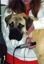
Laser therapy is used to reduce inflammation and pain, promote healing, and reduce adhesions (Figure 1. Laser therapy is used to reduce pain, inflammation, and muscle spasm). The laser is applied directly to the bicipital tendon and the circumference of the glenohumeral joint.
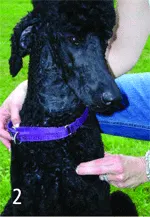
Transverse friction massage is also applied directly to the bicipital tendon; it is delivered in sweeping motions perpendicular to the tendon to realign the fibers of the bicipital tendon and prevent adhesions (Figure 2. Transverse friction or cross-friction massage is applied directly to the bicipital tendon in a sweeping fashion). In acute cases, treatment should be performed for 5 minutes daily. In more chronic cases, treatment may be performed for longer periods in an attempt to recreate acute inflammation. Massage lasting 10 minutes is usually adequate, followed by stretching of the biceps. Neither NSAIDs nor corticosteroids are recommended because the goal is to continue, not interrupt, the inflammatory process.2
Stretching involves flexion of the shoulder combined with elbow extension. Stretching should be held for 15 to 20 seconds and repeated at least 3 times. In acute cases, microtears in the biceps tendon should be prevented by avoiding vigorous stretching.
Pain-free, passive range of motion should be performed at the shoulder and elbow. Attention should also be paid to the scapulothoracic joint; mobilizations may be applied to this area. Therapy should focus on restoring weight-bearing on the affected limb with weight-shifting exercises. There will undoubtedly be weakness in the postural musculature of the shoulder complex, and the atrophy may be controlled with weight-shifting and balance activities.
Balance activities may be performed on a large therapy ball, rocker board, or the ground (Figures 3 and 4).
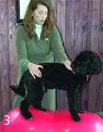
Figure 3. Dog on a PhysioRoll (gymnic.com) for stabilization and balance exercises of the forelimb
Figure 4. Rocker board for stabilization and strengthening of the forelimb
Leash walking is mandatory, and owners must be reminded about the limitation of jumping activities. Once lameness has subsided for at least 7 days, activities may be increased; expanded activities include stepping over cavaletti rails, walking on an underwater treadmill, and progression to jumping.
Additional Rehabilitation
Exercises performed for acute cases should also be performed for chronic cases, with an eye toward increase in activity and return to function.
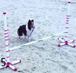
For example, if the dog is involved in agility, a focus on a return to jumping should be part of the rehabilitation program. Low-level plyometrics should be initiated first. Then jumping can be performed on a soft surface, such as soft matting or sand (Figure 5. Begin jumping activities on a soft surface, such as sand). Additional controlled activities, such as ball playing and running, should also be introduced slowly.
At no time during the rehabilitation process should lameness return. Such an occurrence indicates that the shoulder complex is not strong enough to handle the activity and that inflammation and irritation have returned, inhibiting the healing process. If lameness and irritation cannot be controlled, surgical intervention and further exploration of the shoulder complex may be necessary.
Monitoring
Owners should be made aware that monitoring is critical. Lameness is a sign of increased inflammation and should be reported to the rehabilitation practitioner or referring veterinarian. The patient should gradually improve with proper treatment and rehabilitation. Client education to emphasize the need for adherence to controlled exercise is essential. Clients, especially those with performance dogs, are often eager to return the dogs to activity and may push them too quickly.
In General
A multimodal approach to soft tissue injuries of the shoulder, such as bicipital tenosynovitis, is mandatory for a positive outcome. Physical rehabilitation is a strong component of management, and the dog should be referred to a certified rehabilitation practitioner for assistance.
A systematic approach with rehabilitation, proper medical management, and compliance is required. Treatment may last from a few weeks to 6 or 7 months depending on the complexity of the problem and the client's goals for the dog. Performance dogs often require additional rehabilitation and care to reach the goal of returning to performance.
Costs for rehabilitation vary from $50 to $100 a treatment, and treatment frequency may range from a few times a week for several weeks to a few times a month for several months.