Feline Hepatic Lipidosis: From Diagnosis to Treatment
Garret E. Pachtinger, VMD, DACVECC, VETgirl; Veterinary Specialty and Emergency Center, Levittown, Pennsylvania

This article was reviewed in April 2025 to confirm its continued clinical relevance.
Feline hepatic lipidosis is a potentially fatal intrahepatic cholestatic syndrome that develops in cats in association with anorexia (of 2-7 days’ duration) and a catabolic state (ie, a state of negative energy balance). Most affected cats are obese or overweight, middle-aged (median age, 7 years), and/or domestic shorthair breed (see Suggested Reading).
Hepatic lipidosis can occur secondary to another disease process or be a primary, idiopathic disease; anorexia is usually the predisposing factor. Although a simple household stressor may lead to appetite loss, anorexia can result from heart disease, kidney disease, chronic feline lower urinary tract disease, upper respiratory disease, cancer, and pancreatitis.
Clinical Signs
Patients are typically presented with inappetence, lethargy, vomiting, diarrhea, and weight loss. Most cats are alert, but some may exhibit lethargy and depression caused by hepatic encephalopathy or weakness caused by electrolyte derangements (eg, hypokalemia).
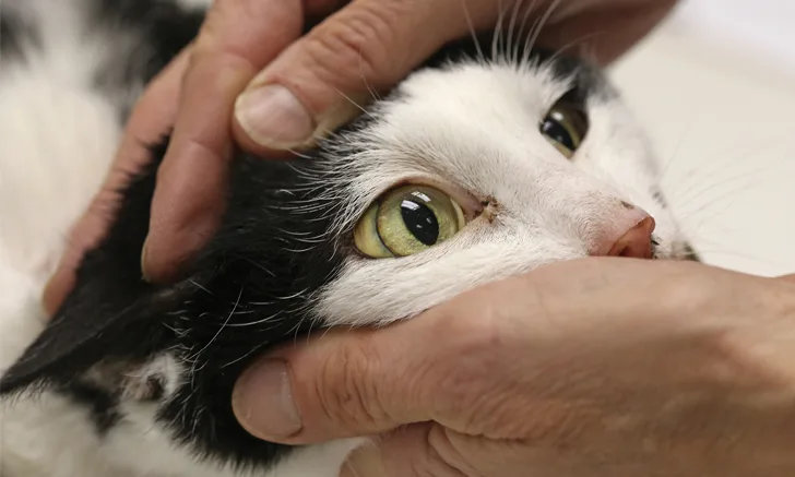
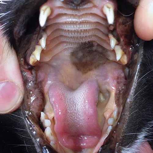
Common examination findings include icterus (Figure 1), signs of dehydration (eg, skin tenting, dry mucous membranes, sunken eyes, dull corneas), an unkempt hair coat, a pendulous abdomen with cranial organomegaly (ie, hepatomegaly), and weight loss often exhibited by dorsal muscle wasting. Specific examination findings may be associated with an inciting, underlying disease process (eg, abdominal pain accompanying pancreatitis, nasal discharge accompanying upper respiratory infection).

Icterus of the hard and soft palate in a cat with hepatic lipidosis
Diagnosis1
How I Diagnose Feline Hepatic Lipidosis
Obtain CBC and serum chemistry profile
Identify electrolyte abnormalities
Pursue abdominal ultrasonography and thoracic radiography
Perform coagulation profile
Pursue cytology and histopathology
CBC findings typically include nonregenerative anemia (Heinz body anemia may also be present) and a stress leukogram, which typically has a characteristic WBC differential (ie, mature neutrophilia, lymphopenia, eosinopenia). Monocytosis is less common in cats than in dogs.
Serum chemistry profile findings primarily reflect cholestasis, including markedly increased alkaline phosphatase and increased serum bilirubin. Transaminases (eg, alanine transaminase) may be slightly elevated, but it is less common for a cat to present with primary hepatic lipidosis and have markedly elevated transaminases and only a mild elevation in alkaline phosphatase. Gamma-glutamyl transferase is often within the normal range (0-10 U/L); elevated levels should prompt concern for other disease processes (eg, pancreatitis). Hypoglycemia is uncommon, as >70% of the functional liver mass must be lost before hypoglycemia can occur (see Suggested Reading).
Electrolyte abnormalities may include hypokalemia and hypophosphatemia; hypomagnesemia is less common. Electrolyte derangements may result from decreased intake and possible GI losses.
Abdominal ultrasound typically reveals liver enlargement (ie, hepatomegaly) with diffuse hyperechoic parenchyma, hyperechoic falciform fat and renal cortices, and an isoechoic spleen. Additional findings may include pancreatitis, triaditis (ie, concurrent cholangitis, pancreatitis, inflammatory bowel disease), biliary disease, and thickening of the small intestinal tract, which may indicate inflammatory bowel disease vs lymphoma.
Thoracic radiography is indicated in older patients or if cardiopulmonary disease is suspected. If a neoplastic process is suspected, 3-view chest films should be obtained.
A coagulation profile, specifically evaluation of prothrombin time and partial thromboplastin time, is an important part of the diagnostic evaluation. Cats can become vitamin K-deficient in <7 days (see Suggested Reading), and, in the author’s experience, >50% of cats with hepatic lipidosis have coagulation abnormalities. Coagulopathic patients may be at increased risk for hemorrhage after invasive diagnostic procedures (eg, aspiration, biopsy). Jugular venipuncture or central line placement also may lead to hemorrhage.
Without cytology, the disease may be suspected but cannot be confirmed. Hepatocellular lipid vacuolation is typically dramatic and diagnostic in feline hepatic lipidosis cases. The author more aggressively recommends this diagnostic approach in older patients to help differentiate ultrasound changes that may also be consistent with lymphoma, as well as for patients in which the history is inconsistent with hepatic lipidosis. Histopathology is rarely used but may be obtained for patients undergoing surgery for an unrelated reason (eg, foreign body, neoplasia).
Treatment
How I Treat Feline Hepatic Lipidosis
Stabilize patient
Initiate enteral support
Restore fluid balance
Correct electrolyte abnormalities
Treat underlying primary disease
Evaluate for and treat coagulopathy, especially vitamin K deficiency
Nutritional support is the cornerstone of treatment for patients with hepatic lipidosis. Interim and emergency intervention includes fluid therapy, assessment and treatment for electrolyte abnormalities, GI support, and appropriate therapy for any underlying disease process.
Enteral Feeding
Enteral feeding should be initiated as soon as possible after fluid balance is restored and electrolyte abnormalities are corrected. It is typically best achieved using a feeding tube; because of the risk for aspiration, patient biting, and development of food aversion, force-feeding with a syringe should be avoided. Patients are often presented for evaluation after several days of anorexia. Administration of an appetite stimulant (eg, mirtazapine, cyproheptadine) rarely induces caloric intake sufficient to reverse hepatic lipidosis and should be used only as a salvage intervention if proper nutritional support has been declined. To determine the appropriate caloric intake, the resting energy requirement (RER; in Kcals) can be calculated using the following equations (see Suggested Reading):
Cats weighing ≥2 kg: 30(BWkg) + 70
Kittens or cats weighing <2 kg: 70(BWkg)0.75
Calculating RER for a 5-kg Cat
RER = 30(BWkg) + 70
RER = 30(5) + 70
RER = 220 Kcal
4 Feedings/Day
Day 1 (25% RER) = 55 Kcal = 14 Kcal/feeding
Day 2 (50% RER) = 110 Kcal = 28 Kcal/feeding
Day 3 and thereafter (100% RER) = 220 Kcal = 55K cal/feeding
The entire calculated RER should not be given on the first day of enteral feeding. Instead, 25% to 33% is typically divided over the first 24 hours (see Suggested Reading). If this is tolerated, the caloric intake is increased to 50% to 67% of RER for day 2; on and past day 3, caloric intake can be increased to 100% of RER. Smaller volumes are preferred initially because prolonged anorexia in patients with hepatic lipidosis may reduce gastric volume and because overfeeding can lead to nausea, abdominal discomfort, and vomiting.
Administration of enteral or parenteral nutrition stimulates the release of insulin, resulting in a dramatic shift of electrolytes from the extracellular to the intracellular space. Electrolytes shifting intracellularly may result in marked hypokalemia and hypophosphatemia; clinical signs include pallor caused by RBC hemolysis, weakness, vomiting, and ventroflexion of the head and neck.
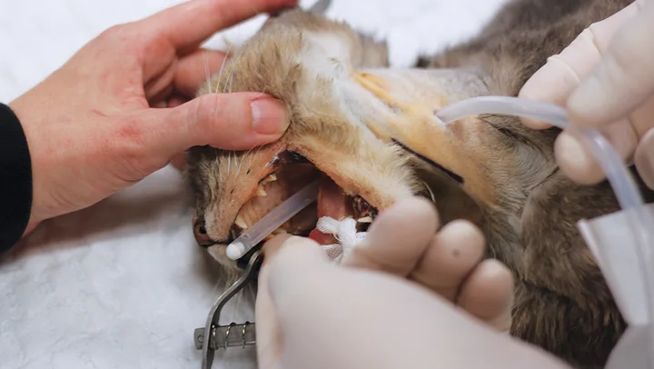
Esophagostomy tubes (E-tubes; Figure 2) are commonly used in patients with feline hepatic lipidosis. Placement requires a stable patient with corrected electrolyte abnormalities, normal cardiovascular status, and tolerance of brief general anesthesia. In the author’s experience, patients are stable for anesthesia and E-tube placement 12 to 24 hours after admission with appropriate hospitalization, treatment, and supportive care. For unstable patients, nasoesophageal or nasogastric tube placement can be considered, as these tubes are often placed without anesthesia. In the author’s experience, E-tubes allow the clinician to provide a more suitable, nonliquid diet (liquid diets often result in diarrhea) with fewer complications than with gastrostomy tubes; they also ease long-term at-home management, as owners can use the E-tube for continued enteral feeding following discharge. After the E-tube is placed, a radiograph should be obtained to ensure appropriate positioning, typically between the seventh and ninth intercostal spaces (Figure 3). Placement in the proximal esophagus may result in tube migration; placement in the stomach may result in esophageal reflux, nausea, discomfort, and vomiting. E-tubes can remain in place as long as needed (in author’s experience, typically 3-8 weeks).
If intermittent E-tube feeding is not tolerated, trickle feeding may be used. Trickle feeding is performed with slow, constant feeding over a longer period of time via syringe pump or fluid pump (Figure 4) that delivers a constant infusion of enteral nutrition through the attached feeding tube.

FIGURE 2
E-tube entering the left midcervical region with the patient in right lateral recumbency. The tip of the catheter can be seen exiting the oral cavity.
Refeeding syndrome is a rare complication that may arise following reintroduction of nutritional therapy after prolonged anorexia; it is characterized by a shift from a catabolic to an anabolic state.
Fluid & Electrolyte Therapy
Fluid and electrolyte therapy is essential for rehydration, maintenance, replacement of ongoing losses, and correction of electrolyte abnormalities and is best provided with a balanced isotonic electrolyte solution. Some clinicians avoid lactate- containing solutions (eg, lactated Ringer solution) because of concerns about decreased lactate clearance associated with hepatic disease. Although this may be a valid concern, intravascular volume replacement is the more immediate concern, and the specific type of balanced isotonic crystalloid is less critical. Cobalamin supplementation can also be considered. In a study evaluating plasma B12 (ie, cobalamin) concentrations in 80 cats with lipidosis, 40% of cats had low cobalamin levels.2
Assessment and correction of electrolyte abnormalities (eg, hypokalemia, hypomagnesemia, hypophosphatemia) are also important. Electrolyte assessment is crucial not only on initial assessment but also once nutrition is reintroduced. See Table 1 for electrolyte support guidelines.
Table 1: Electrolyte Support Guidelines
Ancillary therapy should focus on clinical illness as well as on any diagnosed primary diseases; for example, medications may be given for GI upset manifesting as nausea or vomiting. Vomiting is most commonly addressed with pharmacologic therapy (Table 2).
Table 2: Drugs to Control Vomiting
Cats with hepatic lipidosis are suspected of also having a vitamin K deficiency; thus, when a coagulopathy is diagnosed, vitamin K treatment should be pursued before insertion of feeding tubes, jugular venipuncture, or hepatic aspiration/biopsy. In the author’s experience, most patients supplemented with vitamin K therapy have a corrected coagulation panel (if coagulopathic on presentation) within 12 to 24 hours. This should be documented with a blood coagulation profile (ie, prothrombin time, partial thromboplastin time).
Table 3: Additional Medications to Consider
Conclusion
Successful treatment of feline hepatic lipidosis involves active owner involvement and education. Treatment may require weeks or months of assisted enteral feeding. In the author’s experience, more than 80% of patients can have a full recovery after successful treatment.
Editor's note: This article was originally published in September 2016 as "Feline Hepatic Lipidosis"