Extraction of the Mandibular First Molar in a Dog
Robert Ulbricht, DVM, DAVDC, Associated Veterinary Specialists, St. Louis, Missouri
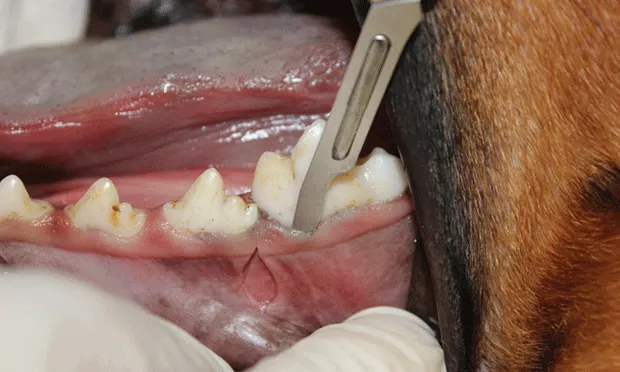
The mandibular first molar, the larger of the two rooted teeth in dogs, is located in the caudal half of the lower jaw. This carnassial tooth functions to break down food with shear force, along with the maxillary carnassial tooth (fourth premolar). This shear force is produced during mastication when the maxillary fourth premolar occludes to the buccal or outside surface of the mandibular first molar, breaking down food for digestion. When a dog bites on a hard object (eg, plastic chew toy, large animal bone) the force may result in a crown fracture.
Complicated crown fractures (ie, those that result in pulp exposure) and advanced periodontal disease are common indications for extraction of the mandibular first molar. Other indications may include dental caries that invade the pulp, root resorption, or location in a mandibular fracture line.
Related Article: Complications of Tooth Extraction: Diagnosis & Treatment
Treatment Options
Treatment options for complicated crown fractures include surgical extraction and endodontic therapy. The most common endodontic therapy performed on the mandibular first molar is standard root canal. If an uncomplicated crown fracture (ie, no pulp exposure) is diagnosed, a dental radiograph is obtained to determine the presence of underlying pathology. In the absence of pathology, rough or sharp fractured surfaces are smoothed (odontoplasty), followed by placement of a sealant on the exposed dentinal tubules.
Surgical extraction is a technique in which the clinician creates a mucoperiosteal flap, removes alveolar bone, and sections the tooth to decrease likelihood of complications. Gentle handling of oral mucosa throughout the procedure minimizes tissue trauma and facilitates rapid healing.
Before extraction is performed, all available treatment options should be discussed with the client. Owner permission is required before extracting any tooth.
The Role of Radiography
Once the patient has been anesthetized for surgery, intraoral radiographs should be obtained to determine the appropriate extraction technique based on the extent of underlying oral pathology. For example, in a patient with marked alveolar bone loss, further removal of alveolar bone may result in a fractured mandible.
Dental radiographs are an essential component of the patient’s medical record for documenting preexisting conditions. Preoperative dental radiographs can also show clients why teeth were extracted and how important future dental care is to manage oral disease.
Complications
Complications that may arise during extraction of the mandibular first molar include mandibular fracture, hemorrhage, alveolar osteitis, fractured alveolar bone that may result in bony sequestrum, excessive force resulting in elevator slippage into the mandibular canal, retained root tip, or root tip entering the mandibular canal.
Taking a surgical approach can decrease the likelihood of complications during the procedure.
Follow-Up
Postoperative care should include appropriate analgesics and antibiotics. Clients should be instructed to feed the patient a soft diet and disallow chew toys for 14 days. Clients should notify the clinician if the dog exhibits any behavioral changes (eg, decreased appetite). A recheck oral examination should be performed 14 to 21 days after surgery.
Step-by-Step: Surgical Extraction of the Mandibular First Molar
What You Will Need
#15 Scalpel blades and handle
Periosteal elevator
Adson tissue forceps
Burs and water-cooled high-speed handpiece (round #2 to #8, cross-cut taper fissure #701L)
Bone curette
Suture material (3-0 chromic gut on a reverse cutting needle)
Needle holders
6-inch Metzenbaum scissors (straight)
Dental elevators or luxators
Extraction forceps
Sterile saline flush
Step 1
After the patient is anesthetized using multimodal pain control protocol (including a mandibular nerve block), obtain an intraoral radiograph to evaluate root structure and mandibular bone integrity.

Step 2
(A) Make 2 full-thickness divergent vertical incisions in an apical direction on the mesial and distal edges of the first molar (black lines). Make the incisions through the gingival and alveolar mucosa to create the full-thickness mucoperiostealflap that will be used later to close the extraction site. Incisions may be extended for further bone exposure if necessary. (B) Once both vertical incisions have been made, make an incision into the gingival sulcus connecting the vertical incisions.


Step 3
(A) Gently slide the periosteal elevator under the attached gingiva at the corner of the flap. Gently rotate and advance the elevator along the attached gingiva until all attached gingiva is freed. (B) Elevate the remaining alveolar mucosa with the same technique in an apical direction.


Step 4
Use a #8 round bur in a water-cooled high-speed handpiece with light pressure to remove the alveolar bone and expose the furcation.
Author Insight
Round burs range in size from #2 to #8, increasing in size with the number. Bur size is selected based on the size of the patient.

Step 5
(A) Section the tooth at the furcation with a #701L bur (cross-cut taper fissure bur) in the water-cooled high-speed handpiece. Place the bur in the furcation perpendicular to the root axis. Once the bur has passed through the alveolar bone, advance it in a coronal direction until it is completely through the crown. (B) To determine complete section of the crown, place a dental elevator between the sectioned segments and rotate it gently. If the 2 segments move independently, the crown has been completely sectioned.


Step 6
Remove the buccal alveolar bone with a #8 round bur in a water-cooled high-speed handpiece. Place light pressure on the bur while moving in a sweeping pattern to each side of the root starting at the marginal bone and moving apically (toward the root tip). Up to 25% of the alveolar bone may be removed initially; removal of 50% may be required.

Step 7
Remove tooth structure on the mesial and distal surfaces (odontoplasty) to allow space for the elevator to slide into the periodontal ligament space. Position the #701L bur parallel to the tooth axis. The mesial and distal portions of the crown may be removed to facilitate extraction by gaining access to the periodontal ligament. Take care not to damage the fourth premolar and second molar teeth.

Step 8
Find the periodontal ligament space and elevate the mesial root (larger of the 2 roots) followed by the distal root. (A) Place a finger stop on the tip of the presharpened elevator/luxator and gently slide it into the periodontal ligament space. Rotate the elevator/ luxator until resistance is met, hold for 15 seconds, then repeat by rotating in the opposite direction. Move the elevator/luxator around the ligament space and repeat until completely around the tooth segment. (B) When the tooth segment becomes mobile, use the extraction forceps to rotate the segment until resistance is met, hold for 15 seconds, then repeat in the opposite direction. Position the jaws of the extraction forceps parallel to the axis of the tooth root. Once the mesial root has been extracted, repeat the procedure for the distal root.
Author Insight
When elevating segments, be careful not to damage adjacent teeth.
Author Insight
Use a short finger stop with the dental or periosteal elevator to minimize trauma if the instrument slips.


Step 9
Obtain a postoperative radiograph to verify that the entire tooth was extracted, and document the condition of the mandible following the extraction.

Step 10
Slightly free the alveolar mucosa from the lingual surface of the extraction site with the periosteal elevator. Place the periosteal elevator between the mucosa and alveolar bone while using a #8 round bur in a water-cooled high-speed handpiece to smooth the sharp edges from the alveolar bone (alveoloplasty). Palpate to ensure no sharp edges remain.

Step 11
Remove debris from the alveoli with a surgical bone curette, and flush with sterile saline.

Step 12
Release tension on the mucoperiosteal flap by applying light pressure on a #15 scalpel blade while incising the periosteum. Make the incision across the base of the flap.
Author Insight
To decrease the likelihood of cutting through the mucoperiosteal flap, use a new scalpel blade and apply very light pressure.

Step 13
Gently place the mucoperiosteal flap over the alveolus. The flap should completely cover the alveoli without tension. Damaged tissue may be trimmed from the margins with 6-inch Metzenbaum scissors (straight).

Step 14
(A) Suture the corners of the flap first with simple interrupted sutures (3-0 chromic gut on a reverse cutting needle). Close between corner sutures by placing the remaining sutures 3 mm apart with 3-mm bites in a simple interrupted pattern. (B) Close vertical incisions in the same manner.

