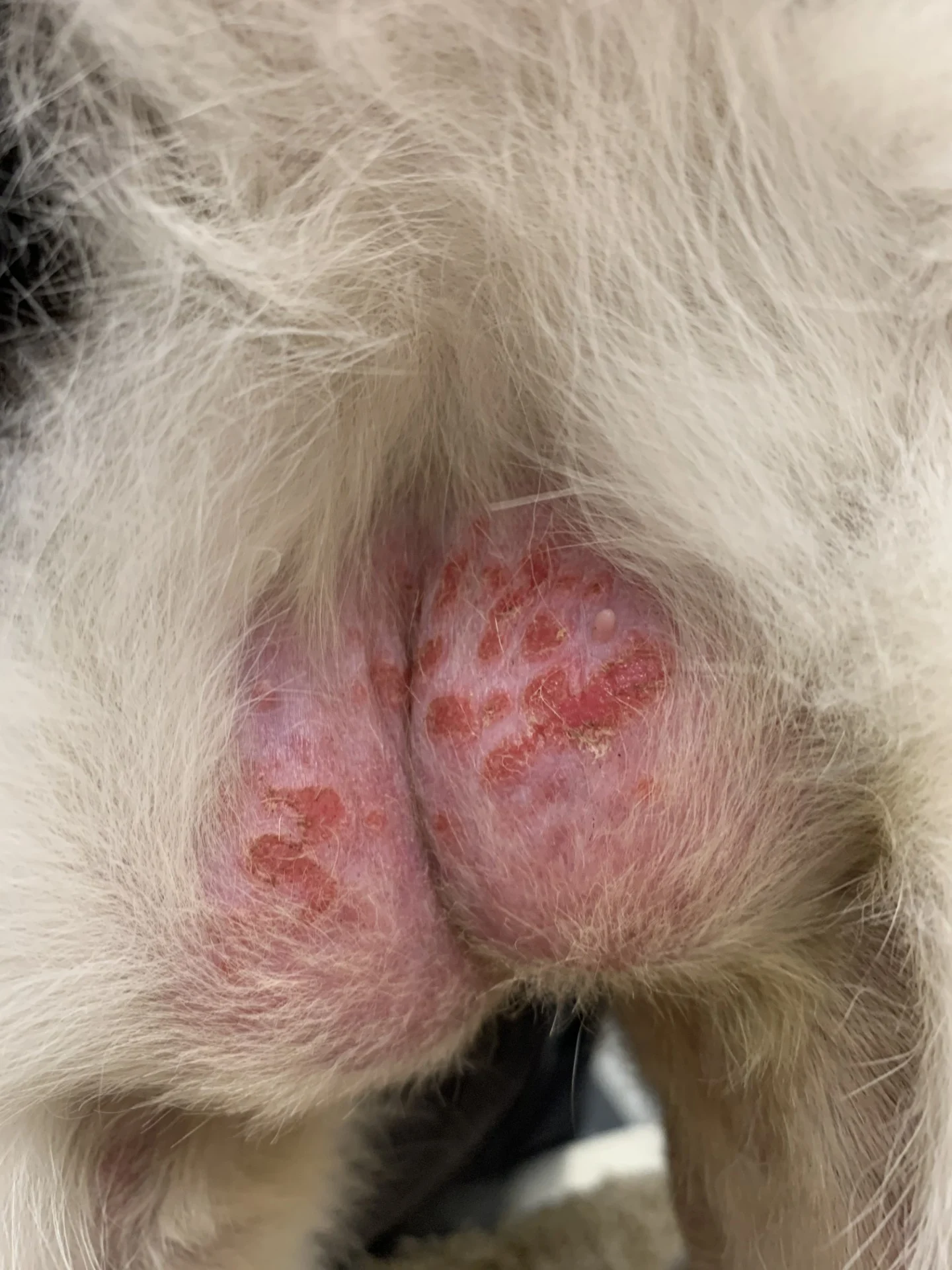
Remi, a 5-year-old, 12-lb (5.4-kg), indoor-only, spayed domestic shorthair cat, is presented for overgrooming and raised, eroded to ulcerated plaques in the abdominal and inguinal regions. Itching, overgrooming, and lesion formation has occurred during spring, summer, and fall months for the past 2 years, with resolution of clinical signs occurring each winter for ≈2 months. Results of a dermatophyte test medium culture performed one year prior to the current examination were negative, and methylprednisolone (20 mg/cat IM) was administered based on a presumptive diagnosis of allergic skin disease. Skin lesions resolved for 6 weeks, but clinical signs gradually reappeared. Remi receives a monthly selamectin/sarolaner topical preventive and is the only animal in the household.
Investigation
On physical examination, BCS is 7/9, thoracic auscultation is unremarkable, and there is near complete alopecia of the ventral abdomen, with rough, barbered hairs on the alopecia margins. Multiple 4- to 8-mm, ovoid to irregular, painful, slightly eroded, and moist plaques are present in the inguinal region (Figure 1). No draining tracts or nodules are present. Dermatologic examination reveals no additional abnormalities, and no fleas are noted. Remi is not reactive to bladder or abdominal palpation.

Multiple annular to ovoid, minimally raised erythematous plaques with mild crusting and self-induced alopecia in the inguinal region of a cat
A skin impression cytology performed on the sores reveals relatively numerous eosinophils (ie, >10 per high-power field), occasional neutrophils (ie, >20 per high-power field), and occasional clusters of cocci bacteria (ie, 5-10 per high-power field; Figure 2).

Impression cytology in an oil immersion. Eosinophils, neutrophils, and cocci bacteria can be seen. 100× magnification