Endotracheal Intubation: Preparation Can Be Lifesaving
Pamela Fettig, DVM, DACVECC, Oradell Animal Hospital, Paramus, New Jersey
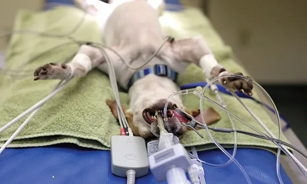
Endotracheal intubation is one of the most important skills a veterinary technician should master. Preparation with the proper equipment and knowledge of the correct technique for intubating every species can be lifesaving.
Indications
General indications for intubation include respiratory and cardiac arrest, general anesthesia, heavy sedation, and any procedure or disease that compromises the airway. Further indications include endotracheal washes, decreased level of consciousness (eg, severe head trauma, brain tumor), periodontal therapy, and upper airway obstructions.1
Equipment & Patient Positioning
Required equipment includes:
3–12 mL syringe to blow up the cuff (depending on the species and tube size)
Endotracheal tubes in varying sizes
Gauze squares (to grab the tongue)
Laryngoscope
Mask
Oxygen
Sterile lubricant
Supplies to secure the endotracheal tube (ie, rolled gauze to tie the tube to the upper jaw, lower jaw, or behind the head).
Patients should typically be placed in sternal recumbency; however, team members should also prepare for and be able to intubate patients in lateral and dorsal recumbency.
Ideally, patients should be pre-oxygenated with an oxygen mask for 3–5 minutes before intubation to allow more time without hypoxemia. Oxygen flow-by should always be available.
Every veterinary technician should have a sound knowledge of the correct intubation equipment and techniques—their patients’ lives may depend on it.
The Procedure
Two team members should be involved in the procedure, with one holding the maxilla with one hand while gently using gauze to pull the tongue with the other hand to give the other team member visual access to place the endotracheal tube. Do not put any fingers in the patient’s mouth and, where applicable, use rolled gauze behind the upper canine teeth to help hold the top jaw open.
Selecting Tube Size
Team members must select the right tube length and placement technique to avoid inadvertent placement into a lung. Various approaches can be used to evaluate proper endotracheal tube size, including palpating the patient’s neck for tracheal size or holding the tube to the nasal septum. With experience, team members will be able to evaluate most patients and select the correct size endotracheal tube, which should be measured from a point immediately rostral to the upper incisors to the thoracic inlet. Ideally, the team member who is intubating the patient should have on hand a tube of the estimated size needed; additional sizes should also be readily available.
Consider safety
The safest endotracheal tubes are the high-volume, low-pressure cuffs because they generate less pressure per given volume of inflation with less risk of tracheal trauma. The cuff should not be inflated by palpating the pilot balloon or according to how “full” it feels because this can lead to overfilling of the cuff or trauma to the trachea. Fill the balloon by listening for air leakage while injecting air and giving the patient a breath.1
Using stylets
Stylets are often recommended for use with small or delicate tubes. Be cautious when using a stylet and ensure it does not protrude from the end of the tube into the trachea, which may result in tracheal trauma, especially if the patient is lightly sedated and coughs or the neck is bent down during intubation.
Proper Placement
To ensure the endotracheal tube has been correctly placed and has not gone into the esophagus, visualize the tube going through the arytenoids and into the trachea (see Figures). Other methods to assure proper placement include observing condensation in clear tubes as the patient breathes, observing the patient’s chest rising, and auscultating lung sounds.
Once correctly positioned, the endotracheal tube should be secured by tying rolled gauze first around the tube and then in a bow behind the upper canines and around the muzzle, making sure it is not too snug. Rolled gauze can also be tied under the tongue and behind the lower canines and lower jaw if placement on the maxilla is not practical.1,2


The anatomy of the oropharynx of the cat (A) and dog (B); the arrows point to the edges of the arytenoids. S indicates the soft palate, E the epiglottis, and V the vocal folds.
Caution
Cats may experience laryngospasm, which may cause the arytenoids to remain closed. Two percent lidocaine can be dropped onto arytenoids via a tuberculin syringe with no needle to help decrease the spasm. Because cats are very sensitive to lidocaine’s toxic adverse effects, use no more than a total of 0.25–0.5 mL;1 1–2 drops are all that is usually needed. Preoxygenation is recommended because a 20–30 second wait is ideal.
Conclusion
In the author’s experience, the best advice is to visualize endotracheal tube placement, which will save time, especially when performing CPCR. Cats can be challenging and having lidocaine available is a must. The ability to intubate in various positions is also an important skill. When moving patients, tubes can become dislodged and may need to be replaced or repositioned mid-procedure.
Every veterinary technician should have a sound knowledge of the correct intubation equipment and techniques—their patients’ lives may depend on it. With practice and experience, these skills can be mastered and endotracheal intubation performed rapidly and efficiently.
This article originally appeared in the March 2015 issue of Veterinary Team Brief.