Elbow Luxation in a Bullmastiff
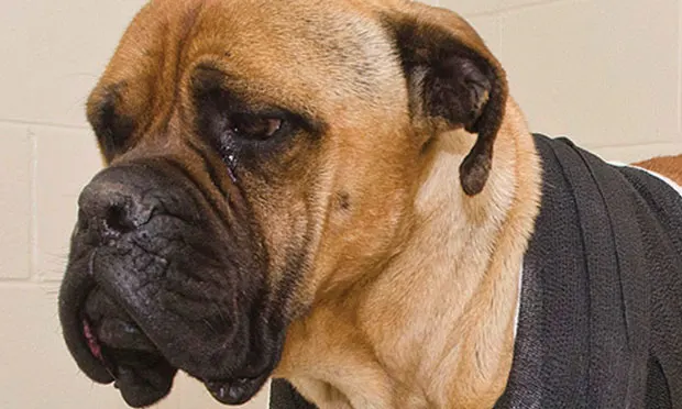
Emma, a 6-year-old spayed bullmastiff, presented for left forelimb lameness of unknown origin.
HISTORYThe owner reported that before Emma was missing for two days, she had appeared healthy. Emma received leftover deracoxib (2 mg/kg PO q24h) that the owner had for a previous right shoulder injury, but Emma did not improve overnight.
The dog was current on vaccinations and had been receiving fish oil (10 mg/kg PO q12-24h) and SC allergen immunotherapy (administered monthly with heartworm and flea/tick preventive) for environmental allergies since 1 year of age.
EXAMINATIONEmma’s body weight was 36.3 kg with a body condition score of 4.5/9. She had marked swelling of her left elbow with decreased range of motion. Crepitus was also appreciated on elbow manipulation. All other findings were within normal limits.
LABORATORY & IMAGING RESULTSCBC, serum biochemistry profile, and urinalysis findings were unremarkable; because of the suspicion of trauma, thoracic and regional films were obtained. Thoracic radiographs were suggestive of mild trauma with mild pneumomediastinum and pneumothorax. Left elbow radiographs revealed left lateral elbow luxation. There was no evidence of fractures.
ASK YOURSELF...
What initial plan is most appropriate for Emma?A. Refer her for a total elbow replacement or arthrodesis.B. Reduce elbow and apply a modified external skeletal fixator.C. Perform open reduction with ligament reconstruction.D. Recommend closed reduction and splinting.
CORRECT ANSWERD. Recommend closed reduction and splinting.
Because of the overall health of the elbow joint and lack of intraarticular fractures, closed reduction should be attempted. More invasive open reductions are saved for chronic luxations or when closed reduction fails. External fixators may also be indicated when orthopedic polytrauma necessitates early weight bearing of the luxated elbow. Salvage procedures (eg, joint replacement, arthrodesis) are reserved for severe degenerative joint disease.
Anesthesia protocols should include pain medications and muscle relaxants to increase comfort and facilitate joint reduction. The radius and ulna usually luxate laterally because the large medial aspect of the humeral condyle generally prevents medial luxation. The goal of closed reduction is to lock the anconeal process into the olecranon fossa and use it as a fulcrum to reduce the elbow. The elbow is flexed ≥90° and the antebrachium pronated and abducted to push the anconeal process over the lateral epicondylar crest into the fossa. Once reduced, the limb is slightly extended with the antebrachium adducted and internally rotated. Pressure should be applied to the proximal radius to force it medially while extending the limb.
ASSESSMENTAfter reduction, the joint should be assessed by examination and radiography for evidence of instability.
The degree of laxity of the reduced elbow should be compared with the normal contralateral limb to assess integrity of the collateral ligaments. Collateral ligament injury is reported in 18% to 50% of elbow luxations, but this may include elongation and complete ligament disruption. This assumption is based on the fact that successful outcome can be obtained with mild to moderate instability following closed reduction.
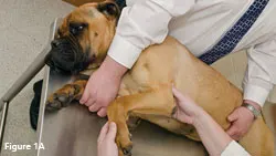
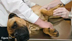
Campbell’s TestIn dogs, pronation of the elbow averages 30° (range, 17°–40°) and supination 50° (range, 31°–67°) with the limb flexed. This should be performed with the carpus and elbow at 90° so the rotation of the antebrachium relies wholly on the collateral ligaments (See Figure 1). This is called Campbell’s test.
(__Figure 1A) When performing Campbell’s test, the carpus and elbow should be held at 90°. (Figure 1B) Supination averages 50° in dogs and stresses the lateral collateral ligament, while (Figure 1C) pronation averages 30° in dogs and stresses the medial collateral ligament.
When measured using a goniometer, normal flexion and extension angles are 36° and 165°, respectively (See Figure 2). If the elbow is easily reluxated with minimal manipulation, open reduction is warranted. If a large decrease in range of motion exists after acute elbow reduction (incomplete or soft tissue entrapment), fractures may be present, indicating further elbow palpation and/or radiography.
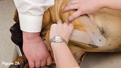
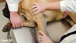
(Figure 2A) Flexion and (Figure 2B) extension angles are measured by placing a goniometer over the neutral point of the joint with the 2 sides parallel on top of the humerus and antebrachial axes.
TREATMENTEmma’s elbow was reduced and deemed stable. She was placed in a spica splint for 2 weeks and after its removal, the owners were instructed to begin gentle range of motion exercises and controlled limb use for 3 weeks. They were also instructed to continue deracoxib and fish oil, and start Emma on Dasuquin (nutramaxlabs.com) according to label instructions for large-breed dogs.
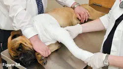
Spica SplintA spica splint should be made to hold the elbow in normal standing position and prevent overflexion. A standing angle of 135° should be used when applying a spica splint to maintain the ulna in contact with the olecranon fossa.
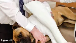
The splint can be made with casting material or a formed aluminum rod. The splint must extend from the toes, over the dorsal midline, to the contralateral side. The splint must immobilize the carpus and shoulder to hold the elbow in place.
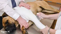
Wrapping the bandage material in front of and behind the contralateral forelimb can help prevent the bandage from slipping back on the chest (See Figure 3). The bandage should not be wrapped around the thorax too tightly.
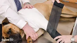
(Figure 3A) A spica splint should contain the traditional layers of a modified Robert-Jones bandage.
(Figure 3B) The reinforcing splinting material must extend from the toes over the dorsal midline to properly immobilize the elbow. (Figure 3C, 3D) The splint should be held firmly to the underlying bandage to ensure a standing angle of 135° to maximize elbow stability.
(
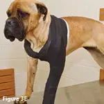
Figure 3E) For added security, the bandage should wrap in front of and behind the contralateral limb.
TAKE-HOME MESSAGES
A complete and thorough physical examination should be performed.
Elbow luxation is usually a result of trauma.
A stable closed reduction of elbow luxation should be splinted for 2–3 weeks with restricted activity for 4–6 weeks.
Avulsion or intraarticular fractures are indications for open reduction and ligament reconstruction.
Owners should be advised of the likelihood of decreased range of motion and preventive measures for arthritis.
FOLLOW-UPNormal bandage care should be advised and includes owner observance at least twice daily to ensure that the bandage is clean and in place and that the toes are not swollen. Once-weekly bandage changes are usually needed unless complications arise. After bandage removal, elbow range of motion should be assessed and physical rehabilitation initiated to optimize range of motion and minimize arthritis.
JENNIFER L. WARDLAW, DVM, MS, DACVS, is assistant professor and chief of small animal surgery at Mississippi State University. Her interests include developmental orthopedic disease, arthritis, physical rehabilitation, and wound care. She has lectured at several national meetings, including the NAVC Conference. Dr. Wardlaw completed her DVM at University of Missouri and a rotating internship and surgical residency at Mississippi State University.
Elbow Luxation in a BullMastiff • Jennifer L. Wardlaw
Suggested ReadingBandaging & splinting canine elbow wounds. Swaim SF, Bohling MW. Clin Brief 11:73-76, 2005.In vitro validation of a technique for assessment of canine and feline elbow joint collateral ligament integrity and description of a new method for collateral ligament prosthetic replacement. Farrell M, Draffan D, Gemmill T, et al. Vet Surg 36:548-556, 2007.Traumatic elbow luxation in 14 dogs and 11 cats. Mitchell KE. Aust Vet J 89:213-216, 2011.