ECG Case Review

Test your knowledge of ECGs and what they tell us with these 5 cases.
Case 1An 11-year-old neutered Labrador retriever presents for an episode of collapse while in the backyard, possibly associated with urination. On physical examination, a slow heart beat is ausculted ~40 bpm.
Answer: Third degree or complete atrioventricular block. The atrial rate is ~150/min and the ventricular rate is ~45/min. The wide, bizarre, and negatively deflected QRS complexes represent a regular ventricular escape rhythm. The P waves have no relationship to the QRS complexes. The PR intervals are not consistent. In some instances, one can visualize the P waves in T wave (arrow). Third degree AV block is considered an urgent rhythm with an elevated risk of sudden cardiac death. Pacemaker implantation is typically recommended if no significant structural heart disease or other significant comorbidities are discovered, as pacemaker implantation is the only intervention shown to improve survival time.1 Atropine response test is generally unrewarding.
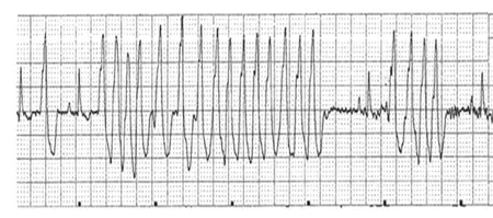
Case 2An 8-year-old spayed boxer presents for an episode of collapse associated with exercise. On physical examination, a rapid and irregular heart beat is ausculted.

Answer: Primarily runs of ventricular tachycardia and a premature complex consistent with arrhythmogenic right ventricular cardiomyopathy (ARVC) of boxers. Two QRS morphologies are seen on this ECG recording. The normal sinus beat (arrow) is contrasted with QRS complexes of the ventricular beats. The run of ventricular tachycardia/flutter at the end of the strip is quite fast, in excess of 300/min (bracket). The wide, bizarre QRS complexes are tall and upright in lead II, suggesting ventricular complexes originating in the right ventricle. This QRS morphology is typical for ventricular arrhythmias associated with Boxer ARVC.
Case 3A 6-year-old castrated large mixed-breed dog presents for lethargy, inappetence, and discomfort. On physical examination, the dog is mildly dyspneic and tachypneic. Heart rate is rapid and irregular.

Answer: Atrial fibrillation with ventricular premature complexes. The rate is fast approximately 220/min. Two distinct QRS morphologies are seen: the narrow positively deflected QRS complexes are those associated with atrial fibrillation (bracket) and the negatively deflected QRS complexes are the ventricular premature complexes (arrow). No distinct P waves are visualized. Small undulations in the baseline suggest fibrillation waves. The rapid irregularly irregular rhythm with no P waves and QRS complexes that have a pretty “normal” appearing morphology are highly suggestive of atrial fibrillation. Atrial fibrillation is usually associated with advanced heart disease and congestive heart failure.
Case 4A 10-year-old castrated German shepherd presents for weakness, lethargy, and anorexia for one day. His heart is difficult to auscult and his pulses are weak. Pale mucous membranes are present, and the heart rate is fast and regular.

Answer: Sinus tachycardia with electrical alternans. The heart rate is ~240 bpm. There is a P-QRS-T wave for every beat but the amplitude of the QRS alternates with every beat. The fast heart rate and alternating short-tall QRS amplitudes are highly suggestive of pericardial effusion and cardiac tamponade, likely due to right atrial hemangiosarcoma in an older German shepherd. An emergency echocardiogram is indicated.
Case 5An apparently healthy 7-year-old spayed beagle is anesthetized for routine dental cleaning. The technician is concerned about the ECG tracing when she first attaches the ECG clips.

Answer: Sinus rhythm with aberrant conduction, likely right bundle branch block. There is a P wave with a consistent PR interval in front of every wide and negatively deflected QRS complex. The wide and negatively deflected QRS complexes are a result of aberrant conduction of the ventricular depolarization wave. A small percentage of dogs have right bundle branch block in the absence of structural heart disease. As long as the blood pressure was normal, one could proceed with the anesthesia event. Nonetheless, one would recommend further evaluation of possible structural heart disease.