Examining the Ear: Otoscopy in Dogs & Cats
Louis N. Gotthelf, DVM, Animal Hospital of Montgomery & Montgomery Pet Skin and Ear Clinic; Montgomery, Alabama
Otoscopic examination of the ear canal is an important part of a regular physical examination, especially because otitis externa is commonly identified in dogs and cats. Ear problems often cause pain that can result in behavior changes. Some neurologic conditions (eg, head tilt, facial nerve palsy, Horner’s syndrome, nystagmus) can develop from otitis externa and otitis media. In dogs, identification of otitis externa can lead to discovery of other dermatologic conditions, as otitis externa is usually secondary to atopic dermatitis and/or food allergies. Aural hematoma can also result from intense scratching and head shaking. In cats, otitis externa secondary to allergy is less likely, but ear mites, middle ear disease, and polyps are often found.
The tympanic membrane can be difficult to visualize and may not be routinely evaluated. Experience gleaned from regular ear examination can help increase awareness of various ear conditions. Video otoscopy provides enlarged, high-resolution images of the ear canal that allow for easier identification of ear conditions.
The following images show ear conditions often identified on video otoscopy. Although these conditions can also be identified on regular otoscopy, video can provide better magnification, lighting, and visibility. Video otoscopes also have the ability to record images for documentation.

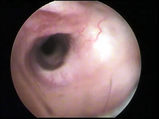
FIGURE 1
A normal ear canal has a glistening surface of scant cerumen coating the epithelial lining. Hairs should be present, and the surface should be pale pink.
Editor’s note: This article was originally published in March 2021 as “Ear Canal Examination.”