Diagnosis of Demodicosis in Dogs & Cats
Karen A. Moriello, DVM, DACVD, University of Wisconsin–Madison
Updated February 2025 by Katherine Doerr, DVM, DACVD; Veterinary Dermatology Center, Maitland and Rockledge, Florida
Profile
Definition
Demodicosis occurs when Demodex spp mites, which are part of the normal flora in dogs and cats, overproliferate in the skin (typically the hair follicle).
Causes
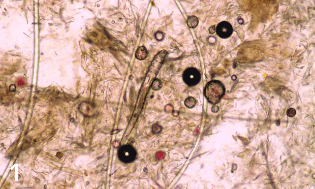
Two species of Demodex spp mites affect dogs: Demodex canis (Figure 1) and Demodex injai (long-bodied mite; Figure 2).1,2
Two species of Demodex spp mites affect cats: Demodex cati (long-bodied mite) and Demodex gatoi (short- and wide-bodied mite; Figure 3).3

FIGURE 1 Demodex canis mite (magnification, 100×)
Signalment
Demodicosis can develop at any age.
No sex predisposition is seen in dogs or cats.
Breeds at highest risk for developing juvenile-onset generalized demodicosis due to D canis include shar-peis, pit bulls, Boston terriers, English bulldogs, French bulldogs, boxers, miniature pinschers, Great Danes, terriers, and pugs.4-6
There is no recognized breed predisposition in cats.
Risk Factors
Risk factors that affect both dogs and cats include chronic severe disease states, neoplasia, long-term glucocorticoid administration, and chemotherapy.
In dogs, risk factors for juvenile-onset generalized demodicosis include breed, pyoderma, coccidiosis, hookworms, short hair coat, and lack of preventive care.4
Localized demodicosis is common in puppies; physiologic stress and debilitation are risk factors.
In cats, D gatoi is contagious. High-density populations have increased risk.
Pathophysiology
Clinical disease results from overproliferation of mites in the skin due to defects or compromise of the skin’s immune system.9
Studies using dog leukocyte antigen class II have identified common markers in young dogs with generalized demodicosis, suggesting this antigen may be an important immunologic risk factor in dogs.7
Demodex spp mites found on skin scrapings, plucked hair, ear swabs, and fecal samples are considered clinically significant when interpreted in conjunction with typical clinical signs.8
Diagnosis
See the Table for a list of clinical forms of demodicosis in dogs and cats.
Table: Demodicosis in Dogs & Cats
Definitive Diagnosis
Dogs
Any finding of mites indicates demodicosis.5,6 Mixed infestations of D canis and D injai can occur.
Mites are found on deep-skin scrapings or via hair trichograms, which are useful for sampling sites close to the eyes or difficult to scrape (eg, interdigital areas).
Hair plucking may be the diagnostic test of choice for sampling dogs with greasy hair.5,6
Cats
In patients with otic demodicosis, mites are found on mineral oil cytology of ear exudates.
D cati mites are usually easy to find on skin scrapings.
D gatoi mites can be difficult to find, even in severely pruritic patients. Suggested tests include wide superficial skin scrapings, hair plucking, fecal flotation (Figure 4), or response to therapy.

FIGURE 4 D gatoi mites found on a fecal sample from a cat with pruritus, self-trauma, and hair loss on the ventral abdomen (original magnification, 400×)
Differential Diagnosis
Canine demodicosis can mimic most skin diseases (eg, hypersensitivity, dermatophytosis, immune-mediated dermatoses, neoplastic dermatoses).
Demodicosis should be considered in cats with hair loss, symmetric alopecia, or pruritus.
Laboratory Testing
Aerobic bacterial cultures of skin should be performed if secondary bacterial dermatitis is present.
CBC, serum chemistry profile, and thyroid profile should be performed in dogs with deep pyoderma that may be septic or dehydrated, in adult-onset demodicosis, and in cats with D cati infestation.
Fecal flotation examination should be performed in cats with pruritus to identify D gatoi.
Genetic testing for multidrug sensitivity gene (MDR1 gene, also known as ABCB1 gene) mutation (also known as ABCB1-1delta) should be performed to screen for drug sensitivity to avermectins in breed-sensitive dogs (eg, herding dogs, sight hounds) and is recommended for dogs with severe generalized demodicosis.3
Impression smears should be performed to diagnose concurrent microbial overgrowth in dogs and cats.
Additional Laboratory Testing
Except for fecal flotation examinations in cats, additional laboratory testing is most helpful when searching for the underlying cause of adult-onset demodicosis in dogs or a medical condition associated with D cati in adult cats. Testing may include but is not limited to:
Blood smear evaluation
CBC
Fecal flotation
Infectious disease titers
Radiography and/or ultrasonography of the thorax and abdomen
Retroviral screening (cats)
Serum chemistry profile
Urinalysis
Fine-needle aspirates of lymph nodes may contain mites.
Skin biopsy with histopathology may be helpful in shar-peis and in dogs with severe pododermatitis.
Read about treatment (including medications), follow-up, complications, and prognosis of demodicosis in Treatment of Demodicosis in Dogs & Cats.