Nasal Planum Disease in Dogs
Darren Berger, DVM, DACVD, Iowa State University

Many pet owners believe a dog’s nose is an indicator of health status and will thus seek veterinary care for any change in coloration or texture or if lesions develop. Following is a discussion of the more common and breed-specific conditions that present with lesions primarily affecting the nasal planum.
Noninflammatory Depigmenting Conditions
Idiopathic Nasal Depigmentation
Idiopathic nasal depigmentation occurs when a nasal planum that was pigmented at birth later loses its color. The condition may wax and wane, but only the nose is affected, with no changes in the nasal planum architecture (ie, the cobblestone-like appearance is preserved (Figure 1). Other noninflammatory depigmenting conditions include snow nose and Dudley nose. Snow nose is a seasonal decrease in nasal pigmentation commonly observed in Arctic breeds and retrievers.1 Dogs with Dudley nose have no nasal pigmentation and are generally affected from birth. Both are cosmetic defects and do not warrant further investigation or therapy.

Idiopathic nasal depigmentation in a 6-year-old neutered male Labrador retriever. Depigmentation is confined to the nasal planum. The cobblestone-like architecture is preserved.
Vitiligo
Vitiligo is a rare acquired disease associated with melanocyte destruction that results in loss of pigment in the hair and skin over several months. The exact pathogenesis is unknown but is thought to be multifactorial, with a hereditary predisposition observed in Belgian Tervuren dogs, rottweilers, and old English sheepdogs.1 Vitiligo is characterized by symmetric macular or patchy leukoderma and leukotrichia that commonly affect multiple areas simultaneously, including the nasal planum (Figure 2). Vitiligo is considered a cosmetic disease in dogs with no consistently successful intervention reported, although spontaneous resolution has been observed.1

Patchy nasal depigmentation caused by vitiligo in a 7-year-old neutered male crossbreed dog
Hyperkeratotic Conditions
Nasal parakeratosis of Labrador retrievers is a familial dermatosis characterized by the accumulation of adherent keratin debris along the dorsal nasal planum with secondary crusting, fissures, erosions, and depigmentation (Figure 3). It typically becomes apparent at an early age (ie, 6-12 months). Lesions may wax and wane but are limited to the nasal planum. Although this is an incurable, lifelong condition, prognosis is good, and early management with frequent application of softening agents (eg, petroleum jelly, propylene glycol) may help prevent severe fissuring.

Significant thick adherent crusting and early fissure formation due to hereditary nasal parakeratosis in a 5-year-old neutered male chocolate Labrador retriever. Image courtesy of A. Kirby, Animal Dermatology Clinic
Nasodigital hyperkeratosis can affect the nose and/or foot pads and is commonly encountered as a congenital hereditary disorder or age-related change. The condition is characterized by an accumulation of thickened, hard, dry keratin along the dorsal nasal planum and edges of the foot pads (Figure 4). Nasal hyperkeratosis may be observed in any breed but is more commonly encountered in cocker spaniels, boxers, and bulldogs. This abnormal accumulation of keratin may lead to pain and discomfort if secondary erosions, ulceration, or fissures develop. Prognosis is good, and treatment should be focused on softening and removing excess keratin. A recent investigation showed daily use of a natural skin-restorative balm to be helpful in managing this condition.2

Idiopathic nasal hyperkeratosis in a 10-year-old spayed American cocker spaniel. Excessive keratin can be seen forming frond- or finger-like projections without signs of inflammation.
Infectious Diseases
Mucocutaneous Pyoderma
Mucocutaneous pyoderma is a bacterial infection that affects the mucocutaneous junctions and nasal planum. Similar to superficial bacterial folliculitis, the infection is normally secondary to an underlying condition (eg, allergic hypersensitivity, endocrinopathy). Lesions may be symmetric and characterized by erythema, crusting, erosions, depigmentation, and discomfort (Figure 5) and are most commonly found along the lip margins (especially the commissures) and nasal planum. Treatment consists of systemic administration of a first-tier antimicrobial (ie, cephalexin, clindamycin, amoxicillin–clavulanic acid) for a minimum of 3 weeks.3 Prognosis is good if the underlying condition can be identified and managed.

Symmetric nasal depigmentation and crusting in a 7-year-old spayed Maltese with mucocutaneous pyoderma
Dermatophytosis
Dermatophytosis (ie, ringworm) may mimic an autoimmune condition such as pemphigus foliaceus, with the nasal planum and/or face solely affected; however, this presentation is uncommon and is typically seen with infections caused by Trichophyton mentagrophytes, which is acquired from direct or indirect contact with rodents. Dermatophytosis is typically characterized by crusting and depigmentation of the nasal planum, along with folliculitis and furunculosis of facial skin when affected (Figure 6). Treatment should include systemic administration of a triazole derivative or terbinafine. Prognosis is good if the point source can be eliminated to prevent recurrence.

Nasal depigmentation and mild crusting secondary to dermatophytosis as a result of an infection with Trichophyton mentagrophytes in a 3-year-old neutered male Saint Bernard. Image courtesy of J.O. Noxon, Iowa State University
Deep Mycotic Infections
Deep mycotic infections (eg, blastomycosis, histoplasmosis) may also present with lesions initially confined to the nasal planum. Depigmentation, swelling, ulceration, and destruction or deformation of the nasal planum are typically present (Figure 7). These conditions have a geographic distribution within the United States, where the infective spores are acquired from the environment via inhalation or inoculation. Prognosis varies from poor to guarded, as many cases have systemic involvement at the time of diagnosis and require extended antifungal treatment regimens with a high occurrence of disease relapse.

Depigmentation, swelling, and ulceration of the right alar fold and nasal planum secondary to an infection with Blastomyces dermatitidis in a 2-year-old neutered male Labrador retriever
Autoimmune Conditions
Pemphigus Foliaceus
Pemphigus foliaceus in dogs occurs as a result of autoantibodies produced against a keratinocyte adhesion molecule that causes cells to detach from one another in the outer layers of the epidermis.4 The disease typically occurs in middle-aged animals, with breed predispositions observed in Akitas, chow chows, Doberman pinchers, dachshunds, and cocker spaniels. The cause of pemphigus foliaceus is idiopathic in most cases, but cases that occur secondary to drug reactions (eg, antimicrobials, topical preventives) or vaccination or as sequelae to a systemic inflammatory condition have been reported.
Lesions consist of pustules, crusts, erosions, and depigmentation and are commonly found along the nasal planum, on the inner aspect of the ears, and on the foot pads (Figure 8).5 Treatment typically involves systemic immunosuppressive medications tapered to the lowest dose required to control disease. Care should be taken not to taper medications too soon or too quickly, as this can lead to clinical relapses and subsequently overall greater cumulative doses. Prognosis varies from guarded to poor and is primarily dependent on the patient’s ability to tolerate the medications.

Nasal depigmentation, ulceration, and crusting as a result of pemphigus foliaceus in an 8-year-old spayed Labrador retriever
Discoid Lupus Erythematosus
Discoid lupus erythematosus is a relatively benign condition. Although the exact pathogenesis of the disease is not known, it is known to be photoaggravated. The nasal planum is the primary location affected, but lesions may also be noted at the periocular, foot pad, perianal, and ear regions. Lesions consist of erythema, depigmentation, loss of the normal nasal planum architecture, erosions, ulceration, and crusting beginning along the dorsal portion of the nasal planum (Figure 9). Treatment protocols vary, with prognosis being fair to good. A key to therapeutic success is limiting or protecting the patient from excessive UV light exposure.

Erythema, depigmentation, loss of nasal architecture, and ulceration along the dorsal aspect of the nasal planum in a 4-year-old spayed Pembroke Welsh corgi with discoid lupus erythematosus
Uveodermatologic Syndrome
Uveodermatologic syndrome is the result of an autoimmune process directed against components of melanocytes. Although the condition may be seen in multiple breeds, it is most commonly reported in Akitas, in which a genetic basis has been proposed.6 The disease process results in granulomatous panuveitis and leukoderma, which may progress to severe crusting and ulcerations (Figure 10); of note, leukoderma may be present in multiple locations and not limited to the nasal planum. Overall, prognosis is fair, and therapy should be primarily focused on the eyes to prevent ocular complications.

Marked crusting, depigmentation, and ulceration of the nasal planum in a 4-year-old neutered male Akita with uveodermatologic syndrome
Nasal Arteritis
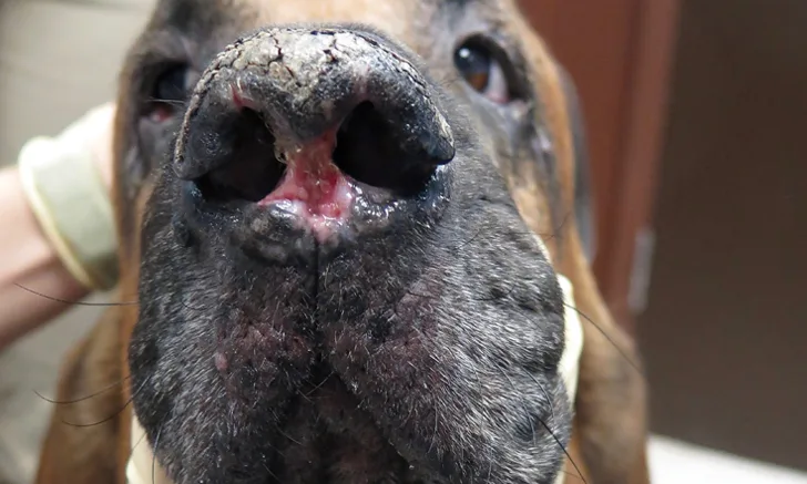
Dermal arteritis of the nasal philtrum is a rare condition most commonly seen in Saint Bernards and characterized by a distinct linear or circular ulcer on the central portion of the nasal planum along the philtrum (Figure 11). The condition can cause recurrent bleeding episodes, which may require immediate intervention. The exact etiology and whether a genetic basis exists are unknown.7 Prognosis is fair, with topical tacrolimus providing an alternative to systemic treatment options.7 Lifelong treatment is required, and permanent scarring in the lesion area is likely.

Significant ulceration of the nasal planum along the nasal philtrum in a 6-year-old neutered male bloodhound with dermal arteritis of the nasal philtrum. Image courtesy of A. Diesel, Texas A&M University
Neoplastic/Infiltrative Conditions
Cutaneous epitheliotropic lymphoma is an uncommon malignancy seen in older dogs. Although the exact etiology is unknown, an association with atopy has been suggested.8 Nasal planum lesions are common, along with depigmentation of mucocutaneous junctions, pronounced erythema, and scaling (Figure 12). Treatment protocols typically involve lomustine +/- radiation therapy; prognosis is poor, with a median survival time of 6 months following diagnosis.9

Nasal planum crusting and depigmentation secondary to epitheliotropic lymphoma in an 8-year-old neutered male English bulldog
Conclusion
Most conditions that affect the nasal planum will present with clinical similarities. Appropriate diagnosis of nasal planum disease requires understanding the conditions that can occur, understanding patient signalment, and ruling out mucocutaneous pyoderma as a primary differential. Once the noninflammatory depigmenting, hyperkeratotic, and infectious conditions have been ruled out, biopsy is typically required for definitive diagnosis. Because it is beyond the scope of this article to thoroughly cover all therapy options for the presented conditions, readers are encouraged to consult additional references (see Suggested Reading) for a more complete discussion regarding disease-specific therapeutic considerations.