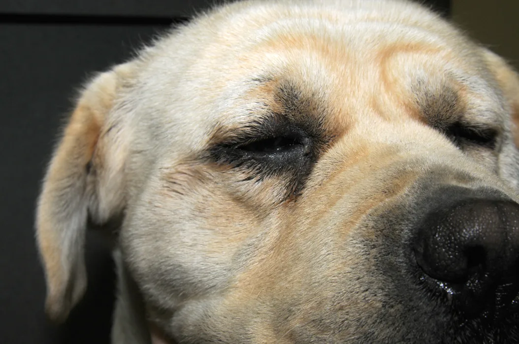
Overview of Canine Hypothyroidism

Updated June 2025 by Andrew Bugbee, DVM, DACVIM; Texas A&M University
Overview
Hypothyroidism is an endocrinopathy caused by decreased production of thyroid hormone. Thyroid hormones are crucial for regulating systemic metabolism and affect all cells in the body. Hypothyroidism therefore affects functions of most organ systems.1 Common clinical signs are related to the dermatologic system and decreased metabolic rate.
Epidemiology
Incidence/Prevalence
Annual prevalence is ≈0.23% (1 in 400 dogs), and lifetime prevalence has been reported to be 1.8% (1 in 55 dogs).2-5
Signalment
Breed Predisposition
Any breed can develop hypothyroidism.
Purebred dogs are at increased risk compared with crossbreed dogs.4-6
A nonexhaustive list of predisposed breeds includes golden retrievers, Doberman pinschers, boxers, schnauzers, Irish setters, and spaniels.2,7,8
Multiple breeds (eg, boxers, Great Danes, Shetland sheepdogs) are predisposed to lymphocytic thyroiditis and are likely predisposed to hypothyroidism.9,10
Age
Mean age at diagnosis is 7 years (range, 0.5-15 years of age).5,7
Likelihood of developing hypothyroidism increases with age.4
Hypothyroidism secondary to lymphocytic thyroiditis appears to develop at a younger age than idiopathic thyroid atrophy.11
Antithyroid autoantibody concentrations are highest in patients 2 to 4 years of age.9
Congenital hypothyroidism is often diagnosed within the first few months of life.
Sex
No sex predisposition has been reported.
Neutered dogs are at increased risk compared with intact dogs.4,7
Genetic Implications
Lymphocytic thyroiditis is heritable in beagles, borzois, and Great Danes; heritability is suspected in other breeds.12-14
Congenital hypothyroidism is an inherited autosomal recessive trait described in several breeds, including rat terriers, toy fox terriers, shih tzus, and giant schnauzers.15-18
Causes & Risk Factors
Primary hypothyroidism (most common) results from autoimmune destruction or idiopathic atrophy of the thyroid glands.
Secondary hypothyroidism is a rarely reported condition caused by decreased thyroid-stimulating hormone (TSH) release from the pituitary gland and is a known component of congenital panhypopituitarism.19
Tertiary hypothyroidism is caused by hypothalamic dysfunction and has not been proven in dogs.
Congenital hypothyroidism commonly results from either thyroid dysgenesis or dyshormonogenesis.
Pathophysiology
Adult-onset hypothyroidism is almost always caused by lymphocytic thyroiditis or idiopathic thyroid atrophy. Lymphocytic thyroiditis is characterized by infiltration of the thyroid gland by lymphocytes, plasma cells, and macrophages. Fibrous connective tissue eventually replaces parenchyma. Idiopathic thyroid atrophy is characterized by thyroid parenchyma being replaced by adipose tissue. Fibrosis and inflammation are minimal. Idiopathic thyroid atrophy may be the end-stage result of thyroiditis. Thyroid carcinoma is a rare cause of hypothyroidism.
Clinical Presentation
Hypothyroidism onset is often insidious, and subtle signs may have been present for months or years prior to diagnosis. Signs are typically seen after 75% of thyroid function has been lost.20,21
Common signs associated with reduced metabolism include weight gain and obesity, lethargy, weakness, cold intolerance or heat-seeking behavior, and mental dullness.
Dermatologic changes are common and occur in up to ≈80% of cases (see Dermatologic Signs).20,21
Diarrhea and constipation occur more commonly in hypothyroid dogs as compared with euthyroid controls.22
Neurologic dysfunction occurs in ≈6% to 29% of cases and often resolves within 2 months following levothyroxine supplementation.19,23
Peripheral neuropathy is the most common manifestation, especially in large breeds, and may result in exercise intolerance, ataxia, weakness, conscious proprioception deficits, or hyporeflexia.
Studies suggest hypothyroid-induced myopathy of the limbs may also directly contribute to these clinical signs.24
Cranial nerve dysfunction (V, VII, VIII) and peripheral and central vestibular disease have been reported.25
CNS signs (eg, seizures, myxedema coma) are rare.
Laryngeal paralysis and megaesophagus have been reported,20,23,26 but no causal relationship has been proven.
Cardiac abnormalities (eg, weak apex beat, bradycardia) are uncommon; however, signs of underlying cardiac disease may worsen with hypothyroidism.19
Reproductive abnormalities include increased periparturient mortality and lower birth weight in puppies born to hypothyroid female dogs and inappropriate galactorrhea in females.27,28
Decreased fertility and cycling abnormalities have not been consistently proven to occur in female dogs with naturally acquired hypothyroidism.
Decreased libido and fertility have been reported in males of other species but were not documented in a study of male dogs with radioiodine-induced hypothyroidism.29
Ocular abnormalities are uncommon and likely associated with the presence of hyperlipidemia. Changes include corneal lipid deposits, lipid-laden aqueous humor, lipemia retinalis, uveitis, secondary glaucoma, and possible reduction in tear production.
Congenital hypothyroidism may result in disproportionate dwarfism, goiter development, or failure to thrive in addition to signs seen with adult-onset disease.
Dermatologic Signs
Bilaterally symmetric and nonpruritic alopecia in areas of increased wear (eg, lateral thorax, flank, tail)—initially sparing the head and extremities (Figures 1 and 2)
Hair may be easily epilated, with absent or slowed regrowth of clipped hair due to hair follicles remaining in the telogen phase of the hair cycle.
Pruritus may be present in dogs with concurrent infectious pyoderma.
Seborrhea (sicca or oleosa), hyperpigmentation, hyperkeratosis, or myxedema (Figures 3 and 4)
Dry, brittle, or dull hair coat
Increased incidence of bacterial pyoderma, otitis externa, folliculitis, generalized demodicosis, and Malassezia spp infections

FIGURE 1
Dog with bilaterally symmetric, nonpruritic, truncal alopecia secondary to hypothyroidism
Differential Diagnoses
Dermatologic abnormalities may be caused by hypercortisolism and alopecia X.
Metabolic signs may be caused by many other disease processes (eg, hypercortisolism, euthyroid sick syndrome, anemia, primary cardiac or neurologic disease).
Find out more in this article, in which 3 internists evaluated dogs prescribed levothyroxine and considered how many were truly likely to have hypothyroidism.
Diagnostics & Diagnostic Findings
Laboratory Findings
CBC, serum chemistry profile, and urinalysis are indicated to rule out other diseases before thyroid-specific tests are conducted.
Most dogs with hypothyroidism have fasting hypercholesterolemia and hypertriglyceridemia.
Mild nonregenerative anemia is common.
Elevated ALP and, possibly, ALT may be present.
Thyroid-Specific Diagnostics
In patients with consistent clinical signs and chemical abnormalities, total thyroxine (tT4) concentration should be measured first.
If the tT4 result is low, TSH concentration and free T4 (fT4) via equilibrium dialysis should be assessed prior to definitive diagnosis.
tT4 assessment is a sensitive but nonspecific screening test.
tT4 values are low in most hypothyroid dogs, but low tT4 alone does not indicate hypothyroidism.
Euthyroid dogs may have a low tT4 due to individual variation, nonthyroidal illness, or drug administration (see Drug Interference With Thyroid Testing).
Euthyroid sighthounds and active working dogs often have tT4 levels below the reference interval.30,31
When present, anti-T4 autoantibodies can cross-react with the tT4 assay and falsely elevate results, which can make euthyroid dogs appear hyperthyroid or hypothyroid dogs appear euthyroid.
fT4 assessed via equilibrium dialysis is a more accurate indicator of thyroid function than tT4.
fT4 is less susceptible to suppression by nonthyroidal illness as compared withtT4.
Equilibrium dialysis methodology excludes the presence of anti-T4 autoantibodies from T4 analysis, preventing antibody cross-reaction.
Measurement of fT4 without equilibrium dialysis offers no diagnostic benefit over tT4 alone.
Due to loss of negative feedback, TSH is often elevated in dogs with hypothyroidism. Increased TSH with decreased tT4 or fT4 is specific for hypothyroidism.
However, up to ≈20% to 40% of dogs with hypothyroidism have a normal TSH concentration.21
A 2 out of 3 rule can assist with thyroid panel interpretation, in which hypothyroidism is likely if 2 of the 3 results suggest this diagnosis.
Low fT4 with an increased TSH concentration is ≈86% accurate for diagnosis.32
Measurement of triiodothyronine (T3) concentrations is less accurate than measurements of tT4 and fT4.
TSH stimulation test evaluates thyroid reserve.
Lack of increase in T4 concentration 4 to 6 hours after TSH administration is standard for diagnosis of hypothyroidism, but the expense of human recombinant TSH limits the test’s clinical utility.
Thyrotropin-releasing hormone stimulation test is less straightforward, less reliable, and more difficult to obtain than the TSH stimulation test.
Thyroglobulin autoantibodies (TgAA) are found in ≈35% to 50% of dogs with hypothyroidism, anti-T3 antibodies are found in ≈10% to 34%, and anti-T4 antibodies are found in ≈6% to 15%.33
Antibody presence is not diagnostic of hypothyroidism; <20% of euthyroid dogs with detectable TgAAs develop hypothyroidism signs within 1 year.34,35
Autoantibodies against T4 may falsely increase tT4 results, while those against T3 may decrease tT3 results on most assays.
Therapeutic Trial
Levothyroxine, a synthetic T4, may be used in patients with consistent clinical signs or biochemical abnormalities, no other significant illness, and/or equivocal test results.
Owners should be instructed to monitor a specific clinical sign or lesion for improvement during the trial.
Some conditions may appear to respond to supplementation in euthyroid dogs, which could delay diagnosis of the actual comorbidity.
If a clinical response is evident, therapy should be discontinued and the patient observed for recurrence. If signs recur, hypothyroidism is confirmed.
Drug Interference With Thyroid Testing
Several medications can affect thyroid hormone concentrations. With the exception of sulfonamides, most drugs rarely cause clinical hypothyroidism.36
Glucocorticoids decrease tT4, fT4, and sometimes TSH in a dose-dependent manner. Performing thyroid testing in dogs receiving corticosteroids should be avoided if possible.
Chronic administration of phenobarbital can cause decreased tT4, fT4, and (less commonly) increased TSH but does not cause clinical hypothyroidism.
Sulfonamides block T3 and T4 synthesis via thyroid peroxidase inhibition, and long-term administration can cause clinical hypothyroidism with increased TSH.
Reversible with drug discontinuation
Of NSAIDs, aspirin is most likely to decrease tT4 and may decrease fT4 with chronic, high-dose administration.
Carprofen, meloxicam, and deracoxib do not appear to alter canine thyroid function.
Toceranib, a tyrosine kinase inhibitor, can increase TSH in the absence of low tT4.37
Potassium bromide does not appear to affect canine thyroid function.
Imaging
Ultrasonography is rarely used but may demonstrate decreased echogenicity and smaller-than-average (volume and cross-sectional area) thyroid lobes in hypothyroid versus euthyroid dogs.
Treatment
Inpatient or Outpatient
Almost all hypothyroid dogs are outpatients. Patients with myxedema coma (rare) should be treated with IV levothyroxine and supportive therapy.
Medical
Oral levothyroxine supplementation is initiated at 0.02 mg/kg every 12 hours. The dose should be based on the dog’s ideal body weight or surface area for larger dogs (0.5 mg/m2). The starting dose should not exceed 0.8 mg total every 12 hours.21
Many patients well-controlled on initial therapy can eventually be transitioned to administration every 24 hours.
Levothyroxine should be given on an empty stomach; administration with food is acceptable but can decrease bioavailability, which may require higher doses.38
Brand name or FDA-approved generic products are recommended, as bioavailability varies among products.
The patient should be maintained on the same product, otherwise dose adjustment and/or additional therapeutic drug monitoring may be needed.
Precautions
Because thyroxine increases cardiac oxygen demand, the initial dose should be decreased by 25% to 50% in patients with cardiomyopathy.19
Treatment of hypothyroidism may precipitate an Addisonian crisis in patients with undiagnosed hypoadrenocorticism.
Before initiating levothyroxine therapy, hypoadrenocorticism should be ruled out with a baseline cortisol and/or ACTH stimulation test in patients with consistent signs (eg, reduced appetite, weight loss, vomiting, diarrhea) or blood work (especially electrolyte) abnormalities.
Treatment at a Glance
Levothyroxine should be started at 0.02 mg/kg PO every 12 hours. The initial total dose should not exceed 0.8 mg every 12 hours.
Doses should be calculated based on the patient’s ideal body weight, and the presence of comorbidities should be considered when determining the safest starting dose.
Ideally, supplementation should be administered on an empty stomach, but this is not required.
Monitoring & Follow-Up
Therapeutic monitoring should begin no earlier than 4 weeks after levothyroxine initiation, unless the patient does not show improvement or develops signs of hyperthyroidism (eg, restlessness, panting, weight loss).
Similar principles apply to monitoring future dose adjustments.
Initial dose determination relies on tT4 levels from blood work performed 4 to 6 hours after levothyroxine was administered (ie, post-pill, peak concentration).
The dose is established when the post-pill tT4 is in the upper half to slightly above a laboratory-established reference interval and a clinical response to therapy is seen.
Once an appropriate dose is established,a post-pill tT4 concentration should be measured every 6 to 12 months in clinically well-controlled dogs.
After 2 to 3 months of good control on administration every 12 hours, tT4 levels from blood work performed at the time levothyroxine is due (ie, pre-pill, trough concentration) can be measured to determine whether transition to administration every 24 hours is appropriate.
The pre-pill tT4 should be in the middle of the laboratory reference interval.
A low pre-pill tT4 suggests administration should remain every 12 hours.
Dogs with a low pre-pill tT4 may still be trialed with administration every 24 hours by increasing the dose by 50% to 100%.
If the dose is increased, another post-pill tT4 should be checked after 4 weeks to confirm peak concentrations remain acceptable.
Dogs initially started on administration every 24 hours should be monitored using both pre-pill and post-pill tT4 concentrations.
Another goal of therapy is to normalize an elevated TSH concentration (if increased at time of diagnosis).
Clinical improvement (eg, improved energy level) is typically noted soon after treatment initiation.
Lack of improvement within 1 to 2 weeks should prompt re-evaluation of diagnosis and/or investigation into presence of other comorbidities.
Weight loss and dermatologic changes improve slowly and may take several months to fully resolve.
Alopecia may initially worsen after treatment initiation due to retained hairs being expelled as hair follicles restart growth cycles; however, signs of new hair growth are typically noted within the first month of treatment.
Neurologic changes may initially improve rapidly, but full resolution can take several months.
Spectrum of Care
Management of hypothyroidism includes tailoring options to match the needs and/or resources of the patient and pet owner. Although high-level care involves definitive diagnostics, screening for other comorbidities, and frequent therapeutic drug monitoring, some owners may decline additional testing beyond a tT4 concentration.
Measurement of fT4 by equilibrium dialysis is often the most expensive aspect of a thyroid panel. A cost-effective alternative is consideration of clinical signs in combination with a tT4 and TSH concentration.
When diagnostic options are limited, a treatment trial based on high clinical suspicion alone may be warranted.
Treatment without appropriate diagnostics should ideally be avoided because the cost of treatment with monitoring is expensive over time, and inappropriate diagnosis may lead to delayed diagnosis or progression of another disease.39
Approaches to monitoring can also be tailored to owner limitations, which may necessitate less frequent drug monitoring and therapeutic goals focused on mitigation of clinical signs.
Being flexible with an approach to management that aligns with owner needs promotes patient well-being through personalized and compassionate care.
Complications
Lack of therapeutic response is common with an incorrect diagnosis (eg, based on low tT4 alone). Other possibilities include decreased GI absorption, poor compliance, and concurrent unidentified skin problems(eg, flea hypersensitivity, atopy).
Prognosis
With proper treatment and monitoring, prognosis is excellent with good quality of life.