Dental Attrition: A Case-by-Case Review
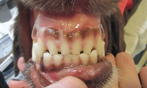
Case 1
Breck, a 6-year-old Labrador retriever, presented for dental cleaning. He enjoys playing fetch and has a collection of balls, particularly tennis balls.
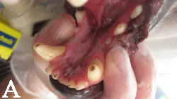
(A) Occlusal view of lower incisors, canines, and premolars. Note the shortened occlusal surfaces with central discoloration. The color change is consistent with reparative dentin formation. Reparative dentin color can range from light tan to reddish-brown to chocolate brown. The surface is typically very smooth. These findings are very common in dogs who favor tennis balls. Over time the abrasive surface of tennis balls will wear away enamel and dentin. If the wear is slow enough and the tooth is still alive, reparative dentin will form. Application of a dentinal sealant is not necessary, since the tooth has already provided its own natural barrier.
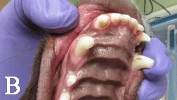
(B) Another example of tennis ball attrition, this time in a younger dog. Occlusal view of upper incisors, canines, and premolars. Note the very smooth worn occlusal surfaces. Tactilely the surfaces are glass smooth, consistent with reparative dentin formation. However, there is very little color change. Sometimes it can be difficult to determine if reparative dentin formation has occurred or is in the process of forming. If dental radiographs appear normal, prophylactic application of a bonded dentinal sealant could be considered.
Related Article: Dental Wear: Managing Attrition & Abrasion
Case 2
Biscuit, an 8-year-old female intact German shepherd dog, presented for assessment of dental wear and multiple tooth fractures. With no history of home dental care, Biscuit was allowed access to bones, which she chewed on frequently.
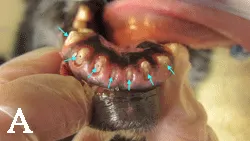
(A) Frontal view of lower incisors showing generalized severe dental wear. Arrows show all teeth with pulp exposure, which indicate that treatment in needed. Extractions were recommended, although root canal therapy could be considered if roots are not resorbing.
(B) Radiographs of lower incisors. Severe dental attrition with pulp exposure at all incisors. Note also the pronounced pulp canal sclerosis, especially for an 8-year-old dog. Canal sclerosis of this degree in a patient of this age is consistent with chronic pulpitis prior to exposure of the canals and tooth death.
Related Article: How to Interpret Dental Radiographs
Case 3
A 2-year-old mixed-breed dog displays dental attrition as a result of orthodontic malocclusion.
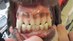
(A) Front view of the incisors. This dog has a mild form of class-3 malocclusion (mandibular mesiocclusion), which has resulted in a level bite (ie, the incisal edges are inappropriately in direct contact).
(B) Lateral view of incisors and canines. Note the edge-to-edge incisor contact, resulting in excessive wear. This dog also constantly carried tennis balls, resulting in excessive wear of the canine teeth.
Case 4
Sienna, a 2-year-old spayed female chow mix, presented for treatment of a fractured left lower canine. Examination revealed tooth-on-tooth wear resulting from orthodontic malocclusion:
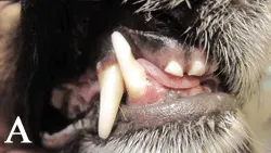
(A) Right front view of canines and incisor. Note the curvilinear groove at the right lower canine (tooth 404) extending from the distolingual to mesiobuccal surfaces. This wear pattern resulted from inappropriate tooth contact with the right upper third incisor (already extracted in this photo).
(B) Lateral view of tooth 404. Although the source of chronic attrition (the right upper third incisor) has been eliminated, the wear pattern has resulted in a structurally weakened tooth, potentially more prone to fracture. At the time of upper incisor extraction, tooth 404 received application of a bonded dentinal sealant. A sealant helps eliminate sensitivity and prevent influx of bacteria via the exposed dentinal tubules. This tooth should be assessed radiographically to help rule out pulpitis or pulp necrosis. Depending on findings, treatment options include careful monitoring with serial radiographic follow-up, root canal treatment (if endodontic disease), and/or restoration to provide strength to the weakened tooth structure.
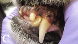
(C) Left front view of canines and incisors. In the same patient the left lower canine (tooth 304) displayed the same wear pattern at tooth 404, a result of inappropriate contact with the left upper third incisor. The structural weakness likely contributed to the fracture of this tooth.
(D) Lateral view of tooth 404 after composite restoration. Other options include application of a prosthetic full coverage crown or placement of an onlay (ie, 3/4 crown).
Case 5
Emmy Lou, a 14-year-old female spayed Labrador retriever, displayed dental findings typical for a senior age dog.
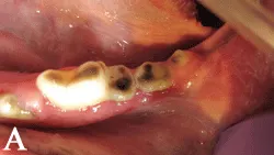
(A) Occlusal view of the left lower molars. Note the flattened occlusal surfaces with chocolate-brown staining. Darker stained reparative dentin is common with older chronic wear. Dental radiography should be performed to rule out endodontic pathology, root resorption, and/or root ankylosis.
(B) Dental radiographs of left lower molars. Despite the crown attrition, these teeth appear in fairly normal health for a dog this age.
Case 6
Gretta, an 11-year-old Labrador retriever, presented for treatment of a fractured right upper fourth premolar. At oral examination, generalized dental attrition was noted at other teeth.
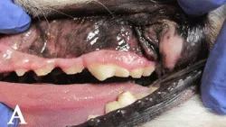
(A) Lateral view of left maxillary premolars. Note the dental attrition, characterized by generalized crown shortening.
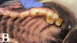
(B) Occlusal view of same teeth. Note the general attrition and color change characteristic of reparative dentin formation. Magnified visual examination and tactile examination with dental explorer revealed no pulp exposure.
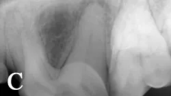
(C) Dental radiograph showing roots of left upper fourth premolar (tooth 208). Note periapical lucency at distal root—a sign of periapical periodontitis (ie, tooth root abscess). Despite the lack of direct pulp exposure and the presence of apparent reparative dentin, this tooth suffered irreversible pulpitis at some point in the past, which then progressed to pulp necrosis, and finally periapical pathology. Treatment is indicated; options include extraction or root canal treatment.
Case 7
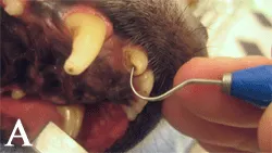
(A) Occlusal view of right upper third incisor. Note how the application of a sharp dental explorer is used to help determine if pulp exposure is present or not.