Deciduous Maxillary Third Premolar Tooth Extraction in a Labrador Retriever
Julie A. Cohen, DVM, The Seeing Eye, Morristown, New Jersey
Ellen A. Scherer, DVM, DAVDC, BluePearl Pet Hospital, Paramus, New Jersey

Uma, a 4-month-old intact female Labrador retriever, was presented for right maxillary swelling of a few days’ duration.
Physical Examination
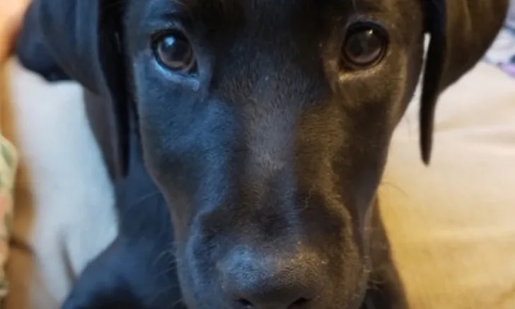
Physical examination was unremarkable except for firm swelling in the right maxillary region (Figure 1).
Diagnosis
Uma was uncooperative and resistant to evaluation of the oral cavity. Examination of the buccal aspect of the right maxillary premolar teeth was unremarkable. A video of the palatal aspect of the right maxillary premolar teeth was obtained, and frame-by-frame evaluation revealed a possible slab fracture of the deciduous right maxillary third premolar tooth (ie, 507; see Suggested Reading for review of modified Triadan system numbering).
Dental radiography via a veterinary digital intraoral sensor and oral examination were performed with Uma under anesthesia. A slab fracture was seen on the palatal surface of 507 with pulpal exposure (Figure 2); a probe was easily inserted into the pulp cavity. No abnormalities were noted on the left side (Figure 3). Radiographs of the fractured tooth and its contralateral counterpart revealed a radiolucent area at the apexes of the mesial (ie, rostral, side of the tooth directed toward the first incisor) roots of 507.

FIGURE 1
Right maxillary swelling
Diagnosis: Fractured Deciduous Right Maxillary Third Premolar Tooth
Treatment & Management
Nerve Block Administration
A right infraorbital nerve block (bupivacaine 5 mg/mL, 0.2 mL) was administered.
Mucogingival Flap Creation
A #15 scalpel blade was used to make 2 full-thickness mucogingival incisions, creating a U-shaped mucogingival flap (Figure 4).1 The distal (ie, caudal) incision was started at the distal aspect of the base of the 507 crown and extended dorsally and distally. The mesial incision was started ≈3 mm mesial to the base of the 507 crown and extended mesiodorsally.
A large L-shaped flap made by one vertical incision at the mesial aspect of the tooth can also be used for extraction of adult maxillary fourth premolar teeth (ie, maxillary carnassial teeth, 108/208).2
The blade tip was inserted into the sulcus around the tooth to release the junctional epithelium at the base of the sulcus on the buccal surface.3,4 The flap was then elevated using a periosteal elevator (Figure 5). Approximately two-thirds of the buccal roots were visible prior to removal of buccal alveolar bone.
Periosteal bone completely covers the roots of adult maxillary fourth premolar teeth.
Initial Tooth Sectioning
A tongue depressor was used to reflect the flap during sectioning, elevating, and tooth extraction. A #2 round ball bur was used with a water-cooled, high-speed hand piece to remove buccal alveolar bone covering the distal tips of the buccal roots (Figure 6). Because deciduous tooth roots are fragile, buccal alveolar bone was removed to expose the entire length of the mesial aspect of the roots. Only 25% to 50% of the root surface needs to be exposed for extraction of adult maxillary fourth premolar teeth.
A C7010 taper fissure bur was used to section the distal root away from the mesial roots following the angle of the mesial aspect of the distal root (Figure 7).
A second cut was made on the distal half of the tooth from the furcation toward the crown, removing a small triangular piece of the crown to facilitate elevation of the distal root and visualization of the furcation between the mesial roots.
Distal Root Extraction
The distal root was elevated by circumferentially advancing an elevator in the periodontal ligament space (between the tooth root and bone), followed by gentle rotation to stretch and fatigue the periodontal ligament, resulting in extraction of the root (Figure 8).
Mesial Root Extractions
A taper fissure bur was used to separate the buccal and palatal mesial roots (Figure 9).
The mesiobuccal root was elevated by circumferentially advancing an elevator in the periodontal ligament space, followed by gentle rotation to stretch and fatigue the periodontal ligament, resulting in extraction of the root (Figure 10).
The remaining mesiopalatal root was elevated by circumferentially advancing an elevator in the periodontal ligament space, followed by gentle rotation to stretch and fatigue the periodontal ligament, resulting in extraction of the root (Figure 11).
Extraction Confirmation & Site Closure
Removed roots were inspected visually, and postextraction dental radiography was performed to confirm there were no remaining root fragments (Figure 12). The area was flushed with a rinse containing chlorhexidine and zinc gluconate, and sharp edges of buccal bone were smoothed with a #2 round ball bur.
Periosteal release was not needed to achieve tension-free closure. The flap was closed with 4-0 monofilament absorbable suture in a simple interrupted pattern (Figure 13).
Postextraction Care
Clindamycin (14.7 mg/kg PO every 12 hours for 14 days) and carprofen (2.44 mg/kg PO every 24 hours for 8 days) were administered postoperatively. Antibiotics are not needed after extraction unless there is evidence of infection or osteomyelitis; in this patient, significant firm maxillary swelling and discolored roots indicated concern for osteomyelitis.
Treatment at a Glance
Dental radiography should be performed to assess the fractured tooth and surrounding teeth, some of which may not be erupted. Surgical approach should be determined based on radiographs and anatomy of deciduous teeth.
Extraction involves creating a surgical flap, obtaining buccal root exposure with a periosteal elevator and minimal drilling of alveolar bone, sectioning the tooth, and gently elevating and extracting individual roots.
Removed sections should be examined closely, and postoperative dental radiography should be performed to confirm complete extraction. Flap should be sutured closed with appropriate absorbable monofilament suture.
Adequate pain management should be provided. Postoperative antibiotics are not necessary unless there is evidence of infection or osteomyelitis.
Prognosis & Outcome
Uma was fed water-soaked dry food and restricted from toys for 2 weeks. The oral incision was healed and maxillary swelling was resolved 2 weeks postoperatively.
Discussion
There are no deciduous first premolar teeth. The anatomy of each deciduous premolar tooth is similar to the permanent tooth that erupts distal to it. Deciduous maxillary third premolar teeth (ie, 507/607) are thus anatomically similar to adult maxillary fourth premolar teeth and have 3 roots.5 Juvenile dogs with deciduous teeth often have some erupting adult teeth (ie, mixed dentition).
The treatment of choice for fractured deciduous teeth with pulpal exposure is extraction, which can prevent periapical infection that can damage underlying permanent tooth buds.3,4 Surgical extraction of a deciduous maxillary third premolar tooth is similar to extraction of an adult maxillary fourth premolar tooth; however, caution is needed because deciduous teeth are close to developing permanent tooth buds, and damage to buds can cause enamel hypoplasia or crown malformation in successor teeth.6 In addition, deciduous teeth have thin enamel walls and can break easily during extraction.7 Mixed dentition and unerupted developing adult dentition visible on dental radiographs can cause confusion and complicate preparation for extraction.
Adult maxillary fourth premolar teeth are commonly extracted in dogs due to fracture. Deciduous maxillary third premolar teeth are not commonly extracted. Caps of deciduous teeth that have fallen off after the tooth roots and associated periodontal ligament have resorbed are more common than full deciduous premolar teeth.8
Take-Home Messages
Compared with adult teeth, deciduous teeth can be more difficult to extract because they have thinner and more delicate roots, thinner dentin layers on roots, and less alveolar bone covering roots.
Underlying developing adult teeth can complicate interpretation of dental radiographs.
Care should be taken to avoid damage to underlying developing adult teeth when elevating and extracting deciduous teeth (especially when extracting deciduous canine teeth).
Each deciduous premolar tooth has similar anatomy to the permanent tooth that erupts distal to it.