Crown Amputation for Tooth Resorption
Jan Bellows, DVM, FAVD, DAVDC, DABVP, All Pets Dental, Weston, Florida
Resorption normally refers to a process in which one part of the body draws in or absorbs another part. Tooth and root resorption involves parts of a tooth being broken down by osteoclasts. Although resorption of internal or external parts of the tooth is possible, the latter is more common, and external root resorption occasionally occurs alongside internal resorption. External tooth resorption often begins on the external surface of the root and progresses inward. In addition to partial hard tissue tooth loss, inflammation of the gingiva is common if the resorption is exposed to the oral cavity.
Common Resorptions
Surface, external inflammatory, and noninflammatory replacement resorption are the most common tooth resorptions in dogs and cats.
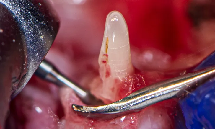
Surface Resorption
Surface resorption may occur secondary to release of osteoclast-activating factors at the site of cementum injury triggered by inflammation. Resorption can occur at any location on the root surface and progress into dentin apically and/or coronally. Surface root resorption (Figure 1) is radiographically characterized by one or more shallow voids that affect the cementum, which can extend into dentin located along the margins of the root. The periodontal ligament space and lamina dura may be locally affected.

Extracted third incisor from a cat with surface external root resorption extending to the oral cavity
When resorption stops, cells from the periodontal ligament proliferate and populate the resorbed area, depositing reparative tissue. Surface resorption is generally self-limiting unless it is exposed to the oral cavity; this is often quite painful because dentin tubules are exposed to heat, cold, and pressure.
External Inflammatory Resorption
External inflammatory resorption begins in the periodontal ligament apical to the cementoenamel junction and at furcations where dentin is exposed. The etiology is unknown. In early stages, odontoclasts resorb and undermine unsupported enamel or cementum, which subsequently breaks away. As the process continues, deeper and more significant amounts of dental tissue are involved. For resorption that begins near the cementoenamel junction and progresses toward the crown, loss of dentin and enamel near the gingival attachment exposes the defect to the oral environment. Inflammation of surrounding tissues occurs, leading to increased sensitivity.
On radiographs, voids (ie, radiolucencies) often extend into the pulp cavity and root canal. With external inflammatory resorption, the tooth roots have similar opacity as surrounding teeth, and periodontal ligament space is visible.
Noninflammatory Replacement Resorption
Noninflammatory tooth replacement of unknown etiology occurs in dogs and cats, in which the root fuses to the bone (ie, dentoalveolar ankylosis), and the tooth is eventually resorbed, becoming part of the alveolar bone remodeling process. This process can take years to complete and is considered a form of healing, as the bone integrates dental hard tissue as part of itself, and the tooth becomes involved in normal skeletal turnover. Osteoblasts form bone in the resorbed area when the resorptive process is complete; thus, dental hard tissues are gradually replaced by bone.
Replacement resorption does not preclude the presence of viable pulp; the pulp can be normal in affected teeth. Lesions sealed below the gingival sulcus may not be painful; however, bacteria can invade the pulp if it is exposed to the oral cavity, leading to painful inflammation. The resorptive process can penetrate the root canal or pulp chamber and, in advanced cases, gradually fill the pulp cavity with new bone. External replacement tooth resorption radiographically appears as a mottled or moth-eaten tooth.
Resorption Classification
Companion animal tooth resorption is often classified by stage and type based on anatomic location and radiographic appearance.1 Stage refers to anatomic loss of dental hard tissue, and type refers to radiographic root opacity and the presence or absence of periodontal ligament space.
Resorption should also be identified as internal or external. Internal resorption takes place in the pulp chamber or root canal and progresses outward; external resorption starts on the outside of the tooth (ie, enamel, cementum) and progresses inward. External resorption should be examined for exposure to the oral cavity and to determine whether roots are significantly replaced with surrounding bone.
STAGES OF TOOTH RESORPTION*
Stage 1: mild dental hard tissue loss (ie, cementum or cementum and enamel)
Stage 2: moderate dental hard tissue loss (ie, cementum or cementum and enamel with loss of dentin that does not extend to the pulp cavity)
Stage 3: deep dental hard tissue loss (ie, cementum or cementum and enamel with loss of dentin that extends to the pulp cavity); most of the tooth’s integrity is retained
Stage 4: extensive dental hard tissue loss (ie, cementum or cementum and enamel with loss of dentin that extends to the pulp cavity); most of the tooth’s integrity is lost
Stage 4a: crown and root are equally affected
Stage 4b: crown is more severely affected than the root
Stage 4c: root is more severely affected than the crown
Stage 5: remnants of dental hard tissue are visible only as irregular radiopacities, and gingival covering is complete
*Adapted from Shope B, Carle D. Tooth resorption in dogs and cats. VetBloom website. February 2, 2017. Accessed December 27, 2021. http://blog.vetbloom.com/dentistry/tooth-resorption-in-dogs-and-cats
Classification by Stage
External tooth resorption stage is a classification system based on affected anatomy. Clinical examination with an explorer and intraoral imaging are used to determine stage (see Stages of Tooth Resorption).
Classification by Type
Tooth resorption type is determined via intraoral radiography based on root opacity and periodontal ligament space (see Types of Tooth Resorption Based on Radiographic Appearance and Figure 2).


Radiograph of a left mandibular molar tooth in a cat with Type 1 resorption (A) in which focal or multifocal radiolucency can be seen with otherwise normal radiopacity and normal periodontal ligament space. Radiograph of mandibular incisors and canine teeth in a cat with Type 2 resorption (B; white arrow) in which narrowing or disappearance of periodontal ligament space is present in at least some areas, and part of the tooth demonstrates decreased radiopacity. Radiograph of the right maxillary third and fourth premolars in a cat with Type 3 resorption (C); the third premolar features Type 1 resorption of the mesial root (arrowhead) and Type 2 resorption of the distal root (dashed arrow) with exposure to the oral cavity.
TYPES OF TOOTH RESORPTION BASED ON RADIOGRAPHIC APPEARANCE*
Type 1 (T1): focal or multifocal radiolucency in a tooth with otherwise normal radiopacity and normal periodontal ligament space
Type 2 (T2): decreased radiopacity in part of a tooth with narrowed or absent periodontal ligament space in at least some areas
Type 3 (T3): features of Type 1 and Type 2 in the same tooth; focal or multifocal radiolucency in a tooth and decreased radiopacity in other areas of the tooth with areas of normal and narrow or lost periodontal ligament space
*Adapted from Shope B, Carle D. Tooth resorption in dogs and cats. VetBloom website. February 2, 2017. Accessed December 27, 2021. http://blog.vetbloom.com/dentistry/tooth-resorption-in-dogs-and-cats
Treatment
Treatment for tooth resorption is largely based on stage, type, and internal/external classification (Table). Extraction is the treatment of choice unless resorption appears to minimally affect the tooth and is confined subgingivally (ie, not exposed to the oral cavity). Intraoral radiography has therapeutic clinical significance for diagnosing patients and creating a treatment plan because only Type 2 resorption should be considered for crown amputation (ie, coronectomy) with gingival closure.
Crown Amputation with Gingival Closure
Bone and cementum‐like tissue eventually replace all or part of the periodontal ligament, dentin, and pulp in teeth with Type 2 resorption. Although complete extraction is the generally accepted treatment for resorption, intentional crown amputation with gingival closure can be effective for cases of Stage 2 to 5 with moderate to advanced Type 2 resorptions or Type 2 resorptions exposed to the oral cavity (ie, external noninflammatory replacement resorption).
Crown amputation with gingival closure is an advanced dental procedure involving creation of a mucogingival flap; use of a water-cooled, high-speed delivery system; and closure without tension that should only be performed after intraoral radiography confirms complete root extraction is not possible based on marked decrease in root opacity and absence of periodontal ligament space. The resorption is sealed off from the oral cavity, and the root continues being replaced by bone.
In teeth affected by Type 3 resorption, roots affected by Type 2 resorption can be treated with crown amputation; the remaining roots should be extracted. Crown amputation typically results in subjectively less trauma and faster healing compared with complete extraction.2
Contraindications for crown amputation with intentional partial root retention include periodontal disease evidenced by horizontal or vertical bone loss, endodontic disease, radiographic presence of a root canal, and chronic gingivostomatitis. Crown amputation should not be performed if intraoral dental radiography is not possible.
Table: Clinical Response to Tooth Resorption Presentations
Step-by-Step: Crown Amputation with Gingival Closure for Radiographically Confirmed Type 2 Tooth
What You Will Need
#11 scalpel blade and blade handle
Fine periosteal elevator
High-speed delivery system
Tapered crosscut fissure bur
#2 round or football diamond bur
Suture holder
4-0 absorbable monofilament or catgut suture on a P-3 needle
Step 1
Identify clinical (A) and radiographic (B) evidence of Type 2 tooth resorption.
Author Insight
This patient has Type 2 resorption of the right mandibular canine with extension into the oral cavity.


Step 2
Use a #11 scalpel blade to make a caudal incision in the gingiva for flap exposure of the coronal root.
Author Insight
For premolars and molars, either an envelope flap or 1 to 2 vertical and sulcular incisions should be made to expose the root.

Step 3
Use a fine periosteal elevator (eg, molt elevator) to separate attached gingiva from the alveolar juga.

Step 4
Use a #2 to #8 sterile round or end‐cutting crosscut fissure bur (A) on a high‐speed, water‐cooled handpiece to remove the tooth crown and 2 to 4 mm of the coronal root apical to the alveolar margin (B).


Step 5
Remove sharp alveolar margin projections with a #2 round or football diamond bur.

Step 6
Perform postamputation intraoral radiography to document the result and confirm all dental hard tissue coronal to the alveolar margin has been removed.
Author Insight
This patient’s left mandibular canine tooth is affected by Stage 5 tooth resorption without exposure to the oral cavity. No treatment is indicated.

Step 7
Draw the mucoperiosteal flap over the alveolus and place single sutures without tension using 4-0 catgut or absorbable monofilament suture.

Step 8
Prescribe anti-inflammatory pain relief medication, and recommend feeding soft food for 2 weeks.
Author Insight
Antimicrobial medication is generally not necessary. The prognosis for complete healing and pain relief is excellent. Surgical site healing and suture dissolution are expected on recheck examination.