Chronic Weight Loss & Diarrhea in a Dog
Micah A. Bishop, DVM, PhD, DACVIM (SAIM), WAVE Veterinary Internal Medicine, Naples, Florida
Clinical History & Signalment
Trixie, a 2-year-old, 44-lb (20-kg) spayed German shepherd crossbreed, was presented for an ≈3-month history of chronic, marked weight loss and small bowel diarrhea. Stool was voluminous, pale in color, and soft and unformed in consistency. Her owner reported that Trixie had a good appetite and appeared to be healthy otherwise. Trial treatment with a hypoallergenic and novel protein diet for 3 weeks did not ameliorate the diarrhea or weight loss.
Physical Examination
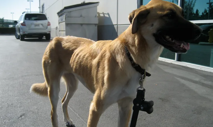
On physical examination, Trixie was bright, alert, and responsive. Vital signs were within normal limits. Her BCS was 2/9 and she had marked muscle wasting (Figure). Abdominal palpation was normal, and soft, yellow feces was detected during rectal examination; flatulence was also noted. The rest of the examination was within normal limits.


Patient showing poor BCS. Image courtesy of Dr. Jörg M. Steiner, Texas A&M University
Diagnosis
Differential diagnoses included intestinal parasitism, chronic enteropathy (eg, food-responsive enteropathy, antibiotic-responsive enteropathy, immunosuppressant-responsive enteropathy), protein-losing enteropathy, juvenile neoplasia, chronic intussusception, chronic foreign body, hypoadrenocorticism (ie, Addison’s disease), chronic kidney disease, chronic liver disease, and infection with Pythium spp, which is endemic in Florida.1
CBC, serum chemistry profile, and urinalysis results were within normal limits. Fecal flotation results were negative.
Because of Trixie’s dramatic weight loss, abdominal radiography and ultrasonography were completed on the day of presentation. Radiographs were unremarkable but revealed mild loss of serosal detail, presumably secondary to patient emaciation. Ultrasound images revealed no mural thickening, abdominal mass, lymphadenopathy, or other abnormality. Adrenal glands were slightly decreased in size.
After Trixie was fasted for 12 hours, serum cobalamin (ie, vitamin B12), folate, canine trypsin-like immunoreactivity (cTLI), and baseline cortisol levels were obtained (Table). The results demonstrated a decreased cTLI, which was diagnostic for exocrine pancreatic insufficiency. Cobalamin was also decreased, which was consistent with ileal pathology. Baseline cortisol was increased, ruling out hypoadrenocorticism.2
Table: GI Panel Results
Diagnosis: Exocrine Pancreatic Insufficiency
Treatment & Long-Term Management
Trixie was initially started on pancreatic enzyme replacement powder at 1 tsp/22 lb (10 kg) of body weight mixed with food.3 She was also given 1 cyanocobalamin tablet daily (1 mg PO every 24 hours is recommended for dogs weighing >44 lb [20 kg]).4 Her owner was instructed to closely monitor Trixie’s stool for improvement in consistency, frequency, and volume and to return to the clinic every 2 weeks for assessment and monitoring for weight gain. Lifelong treatment with enzyme replacement therapy and cyanocobalamin is recommended for exocrine pancreatic insufficiency. Trixie was also empirically dewormed with fenbendazole (50 mg/kg/day for 5 days).5
TREATMENT AT A GLANCE
Pancreatic enzyme replacement therapy is the treatment of choice.8 The dose can typically be tapered over time. These enzyme replacement powders typically contain lipase, amylase, and other proteases.3
Oral cobalamin supplementation can be as effective as parenteral administration, but oral supplementation has not been studied exclusively in EPI patients.9 In the author's experience, the supraphysiologic dose of cobalamin has been sufficient for these patients; however, serum cobalamin concentration levels should be rechecked, especially if there is a lack of response to treatment.
Antibiotic-responsive enteropathy (ie, dysbiosis, small intestinal bacterial overgrowth) is a common complication and may result in partial response to treatment.6,7
Treatment with enzyme replacement is lifelong and expensive. Enteric-coated tablets may be a less expensive alternative, as would be fresh, raw pancreas. Uncoated enzymes can cause gingival bleeding, but this usually can be eliminated by decreasing the dose or administering tablets.6,10
Dietary change is generally not necessary; however, some dogs—especially those with poor response to treatment—may benefit from highly digestible hypoallergenic diets or low-fat diets.6
Eighty percent of dogs respond favorably to therapy, and long-term prognosis is good.6,8
Prognosis & Outcome
Trixie was returned for a recheck examination 2 weeks after presentation. She was rapidly gaining weight, and her stool had improved in quality but was still soft; however, she had also started periodically vomiting daily. Tylosin (25 mg/kg every 12 hours) was given because of her history of low cobalamin in conjunction with the high prevalence of dysbiosis and antibiotic-responsive enteropathy (formerly called small intestinal bacterial overgrowth) associated with exocrine pancreatic insufficiency (EPI).6,7 Dysbiosis was most likely associated with changes in motility, lack of bacteriostatic pancreatic juices, and altered immune function.6 At the next recheck examination, the owner reported that Trixie was thought to be completely back to normal (ie, prior to the development of clinical signs). There were no GI signs, her BCS was 4/9 and expected to continue to improve, and her weight had increased to 57 lb (26 kg). Over the next few months, her BCS returned to normal (ie, 5/9) and her weight increased to 66 lb (30 kg); pancreatic enzyme replacement therapy was tapered to a lower dose. Tylosin was stopped without recurrence of signs ≈6 weeks after diagnosis. Cobalamin supplementation was continued, and Trixie was transitioned to a primary care veterinarian.
TAKE HOME MESSAGES
Although marked weight loss with chronic small-bowel diarrhea and flatulence is a common clinical sign of EPI, it is beneficial to rule out EPI in any patient with weight loss regardless of GI signs.
cTLI is a highly sensitive and specific test for EPI that should always be done on a fasted blood sample.11
Previous administration of pancreatic enzymes does not interfere with cTLI testing.12
Cobalamin and folate derangements are common secondary findings that should be addressed. In a study, low cobalamin was associated with decreased survival.12 Eighty-two percent of dogs with EPI have decreased serum cobalamin concentrations; however, there is some disagreement as to what the cutoff reference interval should be and whether serum methylmalonic acid concentration should instead be assessed, as this may indicate earlier deficiency.13,14
Laboratory findings, radiography, and ultrasonography can be used to rule out common differential diagnoses.
German shepherd dogs and rough-coated collies are predisposed to EPI, likely due to an autosomal-recessive inheritance pattern.15