Cataracts in Dogs
Laura Mancuso, VMD, University of Tennessee
Diane Van Horn Hendrix, DVM, DACVO, University of Tennessee, Knoxville
How do I approach a suspected cataract in a canine patient?
Overview1
Cataracts may occur at any age and in any location in the lens. Location may predict progression risk. Cataracts partially or completely block tapetal reflection and fundic examination and are often classified by stage of maturation and cause.
Stages of Maturation
Incipient (Figure 1): <15% lens volume. Tapetal reflection is minimally obstructed. Visual deficits are not apparent.
Immature (Figure 2): 15% to 99% lens volume. Tapetal reflection is still visible but varies with the degree of cataractous lens. Visual impairment is variable, from minimal to near-complete blindness. Note that partial tapetal reflection and vision may be restored during cortical resorption in hypermature cataracts. This can confound the distinction between hypermature and immature cataracts.
Mature (Figure 3): 100% lens volume with no resorption. No tapetal reflection is visible. Eyes are blind but retain dazzle reflex and pupillary light reflexes (PLRs) if the retina is functional.
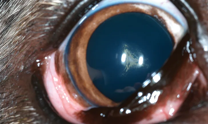
Hypermature (Figure 4): Resorption is present and produces a wrinkled anterior lens capsule with white plaques and multifocal sparkling. Phacolytic uveitis is common.
Morgagnian: Resorption and liquefaction of lens cortex with ventrally positioned (ie, dependent) nucleus. Vision may return.
Trusted content.
Tailored to you.
For free.
Create an account for free.
Want free access to the #1 publication for diagnostic and treatment information? Create a free account to read full articles and access web-exclusive content on cliniciansbrief.com.