Cat With a Cloudy Eye
Randi Rose Sansom, BS, University of Tennessee
Thomas Chen, DVM, DACVO, University of Tennessee

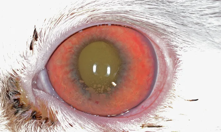
Affected left eye.
Mild conjunctival hyperemia is present. The cornea has 360-degree superficial neovascularization that extends to the axial cornea and is mildly edematous. Throughout the cornea are multifocal-to-coalescing white-gray opacities that are most dense ventrally, consistent with keratic precipitates. The iris appears hyperemic with abnormal vascularization (rubeosis iridis). The pupil is midrange in size, and what can be visualized of the lens appears normal.
THE CASE
Norris, a 10-year-old, neutered male domestic shorthair cat, is presented with a 3-week history of cloudiness and redness of the left eye and inappropriate urination of 2 months duration. He is up-to-date on FVRCP, feline leukemia virus (FeLV), and rabies vaccinations and receives monthly topical heartworm, flea, hookworm, and roundworm preventive. Norris spends time both indoors and outdoors; he has no recent travel history.
On physical examination, Norris is quiet and alert and has a BCS of 3/9. Temperature, heart rate, and respiratory rate are normal. A tense abdomen prevents identification of individual organs on palpation. Seborrhea sicca and a dull hair coat are also noted. No neurologic deficits are observed.
The left eye has an intact menace response but an absent direct PLR and intact left-to-right PLR.
On ophthalmic examination, the right eye appears normal with an intact menace response and intact direct pupillary light reflexes (PLR) but absent right-to-left PLR. Dazzle reflexes are intact for both eyes. The left eye has an intact menace response but an absent direct PLR and intact left-to-right PLR. There is 2+ aqueous flare, conjunctival hyperemia, and episcleral injection, as well as corneal neovascularization and keratic precipitates ventrally (Figure 1). The iris appears hyperemic and vascularized. The lens is only partially visible, but a tapetal reflex is noted through the pupil, and no cataract is present. The flare and corneal changes make viewing the retina difficult. Fluorescein stains of both eyes are negative, and Schirmer tear test results are normal. Tonometry measurements are 13 mm Hg and 8 mm Hg for the right and left eye, respectively (normal, 15-25 mm Hg). No foreign body is noted behind the third eyelid of the left eye. A morphologic diagnosis of anterior uveitis in the left eye is made.
CBC, serum chemistry profile, and urinalysis, along with FeLV and feline immunodeficiency virus (FIV) testing, are recommended. Norris resists multiple attempts to obtain a urine sample, so the owners are given nonabsorbent cat litter for at-home urine collection. The FeLV/FIV SNAP (idexx.com) test is negative. Other results are shown in Table 1.
TABLE 1: CBC & Serum Chemistry Profile Results
*Value is outside the reference range.
FeLV = feline leukemia virus, FIV = feline immunodeficiency virus, IOP = intraocular pressure, PLR = pupillary light reflexes