The Case: The “Complicated” Blocked Cat

Summary
A 1-year-old neutered male indoor cat was presented for lethargy and inappetence of 3 days’ duration. He had not exhibited vomiting or diarrhea, and the owners did not appreciate any changes to his urinary habits. The patient was quiet, alert, and responsive. A large, turgid, painful bladder was detected by abdominal palpation and could not be expressed with gentle pressure. The extruded penis was erythematous with a hard plug palpable in the distal urethra. The patient was anesthetized and the obstruction was alleviated by passage of a tomcat urinary catheter, although removing the blockage was challenging. The tomcat catheter was removed, and a 3.5 Fr 14-cm rigid polytetrafluroethylene-coated (Slippery Sam tomcat) urinary catheter was passed into the urethra with minimal difficulty and maintained as an indwelling catheter. Approximately 70 milliliters of hemorrhagic urine was removed from the bladder, and a sample was collected for urinalysis. The urinary bladder was flushed with saline. Recovery from anesthesia was uneventful. The patient was admitted to the ICU, started on telemetry, and maintained on IV fluids, buprenorphine, and prazosin. Urinary output and catheter patency were closely monitored.
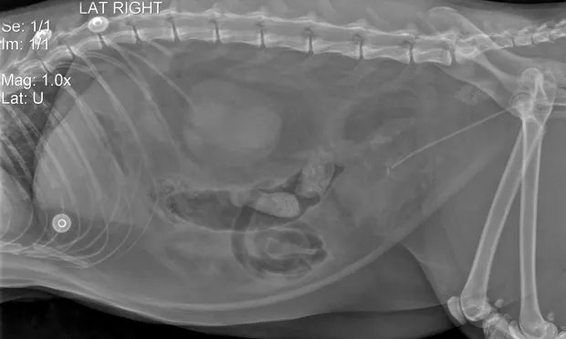
A radiograph immediately after unblocking showed the urinary catheter appeared to be in place at the center of the bladder with no radiopaque urinary calculi noted. The bladder was small, and there was a slight loss of detail in the caudal abdomen, suggestive of effusion (Image 1). Due to ensuing persistent hyperkalemia, the urinary catheter was examined thoroughly. It appeared patent, although urine was not flowing as readily as would be expected. The catheter was flushed, and urine flow improved. An abdominal focused assessment with sonography for trauma (FAST) scan showed a scant amount of free fluid in the abdomen, with a small, intact urinary bladder. Urine output was calculated to be 2.5 mL/kg/hr. The patient remained bright and appeared to be resting comfortably.
About 5 hours later, the patient appeared quiet and more uncomfortable, and it was noted that his urine output had decreased to less than 2 mL/kg/hr; he had gained 0.3 kg since admittance. Repeat abdominal FAST scan showed an increased amount of peritoneal effusion. A contrast cystogram was performed and confirmed uroabdomen, with leakage of contrast at the level of the pelvic urethra (Image 2).
What went wrong and how could management have been improved?
Diagnostic Work Up
Initial Physical Examination Findings
The cat had a historical heart murmur but no other significant health history. The extruded penis was erythematous with a hard plug palpable in the distal urethra.
Hydration: 6%–7% dehydration
Temperature: 98.6⁰F
Pulse: 180 bpm
Respiration: 60 bpm
Systolic blood pressure (Doppler): 140 mmHg
Weight: 6.2 kg, BCS 5/9
Initial Diagnostics
Sodium: 136 mmol/L (range, 147–162)
Potassium: 8.9 mmol/L (range, 2.9–4.2)
Blood urea nitrogen: >140 mg/dL (range, 15–34)
pH: 7.151 (range, 7.25–7.40)
Creatinine: 8.5 mg/dL (range, 0.8–2.4)
Treatment
Anesthesia: IV buprenorphine and midazolam premedication; propofol induction. After intubation, isoflurane was used to maintain a stable plane of anesthesia.
Fluids: 50-mL crystalloid bolus at start of procedure, then 30 mL/hr during procedure. A second 50-mL bolus was administered at end of the procedure.
Cardioprotection: 3 mL 10% calcium gluconate as a cardioprotectant (hyperkalemia), with 6-mL bolus 25% dextrose at start of procedure to decrease serum potassium level.
Continuous electrocardiography: normal rate/rhythm; abnormally tall T-waves
Urethral procedure: A tomcat catheter was passed into the distal urethra and there was an initial firm obstruction 5 millimeters proximal to the tip of the penis. After repeated flushing with sterile saline, the catheter was able to be advanced. There was a second area of obstruction at the level of the pelvic urethra, which eventually yielded to repeated saline flushes and gentle advancement of the catheter. The tomcat catheter was removed, and a 3.5 Fr 14-cm Slippery Sam urinary catheter was passed into the urethra with minimal difficulty and sutured in place as an indwelling catheter. Approximately 70 milliliters of hemorrhagic urine was removed from the bladder, and a sample was collected for urinalysis. The urinary bladder was flushed with saline.
Postprocedure treatment plan:
Plasmalyte*: 25 mL/hr with 5% dextrose
Buprenorphine: 0.09 mg q8h IV
Prazosin: 0.5 mg q12h PO
Urinary catheter care: q6h
Urine output quantification: q4h
*A multiple electrolyte solution
Diagnostics & Postprocedure Management
Due to persistent hyperkalemia, the urinary catheter was examined thoroughly. It appeared patent, but urine was not flowing as readily as would be expected. The catheter was flushed, with subsequent improved urine flow. To address hyperkalemia:
1 unit regular insulin administered IV
2 3-mL boluses 10% calcium gluconate administered IV
5 ½ hours after catheterization: patient remained bright and appeared to be resting comfortably.
Abdominal focused assessment with sonography for trauma (FAST) scan: scant free fluid in abdomen; small, intact urinary bladder
Urine output (calculated): 2.5 mL/kg/hr
10 hours postcatheterization: Cat appeared quieter, more uncomfortable. It was noted that his urine output had decreased to less than 2 mL/kg/hr. He was reweighed and had gained 0.3 kg since admittance. Repeat abdominal FAST scan showed an increased amount of peritoneal effusion.
Diagnostic abdominocentesis:
Creatinine: 8.4 mg/dL (compared to 6.7 mg/dL in serum)
Potassium: 9.9 mmol/L (compared to 6.5 mg/dL in serum)
Contrast cystogram: confirmed uroabdomen, with leakage of contrast at the level of the pelvic urethra (Image 2)
Outcome
Due to the location of the urethral tear, there was concern that the site would not be accessible for surgical correction. The patient was initially managed by leaving the urinary catheter in place and more aggressive fluid diuresis, in hopes that he could be supported while giving his urethral tear time to heal. However, the cat’s hyperkalemia did not resolve, and he became increasingly azotemic, so a peritoneal drain was placed and peritoneal lavage was instituted: azotemia improved. A perineal urethrostomy was performed 2 days after initial presentation. The cat has done well at home since discharge.
The Generalist’s Opinion
Barak Benaryeh, DVM, DABVP
This case is a good reminder of potential complications involved in urinary obstructions. There does not appear to be a good study on how often urethral tears occur, but they are not unique to this case. The clinicians involved did a good job of recognizing and treating the urethral damage.
Initial Catheterization
A tomcat catheter was used initially. Tomcat catheters come in two types: open and closed, and it was not specified which was used here. The advantage to the open style is the ability to flush straight forward without putting pressure on the walls of the urethra, but slightly more trauma may be induced than with a closed catheter during passage, as the latter has a rounded end. Catheters such as the Slippery Sam (or any catheter with a wire) can be problematic as the guide wire can be damaging. We do not know whether the damage occurred to the urethra during passage of the tomcat catheter or the Slippery Sam. There are many methods and tricks of the trade that different clinicians use for obstructed cats. Whatever the method, always be sure to flush the urethra rather than force the catheter forward. Pull the prepuce caudally once the catheter is in the penis; this step straightens the urethral flexure, which can be a point of rupture.
Electrolyte Disturbances
To guard against complications associated with elevated potassium, calcium gluconate was administered, along with dextrose. Calcium gluconate is generally the drug of first choice in hyperkalemia, as it starts to lower potassium immediately. Dextrose is frequently given along with insulin. The insulin was introduced later during the treatment process. Electrocardiography (ECG) was conducted throughout, which is ideal in cases of hyperkalemia. Recognizing the classic ECG changes that occur during hyperkalemia is important, as animals can exhibit such changes at various levels of potassium.
Electrolyte aberrations generally do not occur in isolation: If there is a severe change in one parameter, there are usually changes in several parameters.1 Be sure to monitor blood values throughout treatment. The doctors in this case did an excellent job of staying on top of the electrolyte values and were able to recognize that there was a problem.
Prazosin
Prazosin, an alpha-1 blocking agent, reduces sympathetic tone and is frequently used in obstructed cats to treat urethral spasm and decrease urethral sphincter tone. While it was used in this case and many clinicians, including myself, use this drug, it is worth mentioning that studies have shown conflicting results as to its effectiveness.2,3
Managing Urethral Tears
Many urethral tears can heal without surgical intervention. If a patent catheter can be effectively passed into the bladder and left in place, conservative management is an option. This particular case required a urethrostomy as dictated by the continued decline in clinical status. In the end, a high level of veterinary care and good monitoring turned what could have been a fatal outcome into one that was a success.
Barak Benaryeh, DVM, DABVP, is the owner of Spicewood Springs Animal Hospital. He graduated from University of California–Davis School of Veterinary Medicine in 1997 and completed an internship in Small Animal Medicine, Surgery, and Emergency at University of Pennsylvania. Dr. Benaryeh has also taught practical coursework to first-year veterinary students and was a primary veterinary surgeon for the Helping Hands Program, which trains assistance monkeys for quadriplegic people. Dr. Benaryeh is certified by the American Board of Veterinary Practitioners in Canine and Feline Practice.
The Specialist’s Opinion
Gretchen Statz, DVM, DACVECC
Urethral obstruction (UO) in male cats is common and often frustrating. Complications include severe metabolic derangements (hyperkalemia, azotemia, acidosis), postobstructive diuresis (POD), and urethral tears secondary to urethral catheterization. Recurrence rates are relatively high (23% to 36%)1,2 making it all the more difficult. This case was handled well for the most part.
Fluid Therapy in Urethral Obstructions
Cats with urethral obstruction often have marked hyperkalemia, azotemia, and acidosis. Relieving the obstruction is a priority in reversing these derangements, but fluid therapy also plays an important role. Sodium chloride (0.9% NaCl) does not contain potassium and has been used historically. A balanced electrolyte solution, which does contain some potassium, was used in this case. Despite what logic might dictate, this was a good choice. Studies have shown that potassium-containing fluids (lactated Ringers [LRS] specifically) are safe and effective in cases of UO. In one study, both fluid types (NaCl and LRS) were found to be safe; however, LRS was more efficient in restoring the acid–base and electrolyte balance in severely decompensated cats with UO.3
Fluid “ins and outs” and electrolytes must be monitored closely as POD and hypokalemia can occur after the obstruction is relieved. Fluid rates may be several multiples of normal maintenance rates to compensate for obligate renal losses. Similarly, potassium often needs to be supplemented during the POD phase, even for patients that were initially hyperkalemic. The urine output in this case was lower than the input (17.25 mL/hr out and 25 mL/hr in) with a persistently elevated potassium, which is not typical for postobstruction patients. This disparity and persistent hyperkalemia did prompt reevaluation of the catheter’s patency and eventually ultrasound examination of the abdomen.
The Unblocking Procedure
Urethral plugs can be deeply lodged and pressure from a full urinary bladder can make dislodgement difficult or impossible. It is easy to exert excessive force, especially when a rigid catheter such as a polypropylene (tomcat) catheter is used. The unblocking should occur from flushing the plug back into the bladder with saline and not from advancing the catheter itself. Softer options such as red rubber or polytetrafluroethylene (PTFE or Slippery Sam) catheters can help to avoid excessive force from the catheter. Over-the-needle catheters or lacrimal needles have also been recommended.4,5 In difficult cases, decompressive cystocentesis using a small gauge needle or over-the-needle catheter can help to reduce the pressure behind the urethral plug and allow for easier dislodgement.
Urethral Tears
Urethral tears are an unfortunate complication of urethral catheterization. In one study, the most common cause of urethral tears in cats was trauma associated with urethral obstruction and catheterization.6 In another study, vehicular trauma was the leading cause with urethral catheterization a close second.7 Urethral tears will often heal on their own if a catheter can be left in place while the tear is allowed to heal (5–14 days).8
If urethral catheterization is difficult after a urethral tear is diagnosed, percutaneous antegrade urethral catheterization via fluoroscopy can be used.9 If conservative management with an indwelling catheter is not possible or not successful, then surgical repair is indicated.
Gretchen Statz, DVM, DACVECC, is an internal medicine consultant for Antech Diagnostics. A graduate of University of Wisconsin–Madison, Dr. Statz interned at VCA West Los Angeles and then worked for several years at two emergency/referral hospitals in the Boston area. After completing a residency at VCA Veterinary Referral Associates in Gaithersburg, Maryland, she became boarded in emergency and critical care. Having a strong interest in internal medicine, she has been practicing in that field for the past several years.