In the Literature
Costanza D, Greco A, Piantedosi D, et al. The heart to single vertebra ratio: a new objective method for radiographic assessment of cardiac silhouette size in dogs. Vet Radiol Ultrasound. 2023;64(3):378-384. doi:10.1111/vru.13201
The Research …
Vertebral heart scale (VHS) is widely used in clinical practice as an objective method to assess cardiac size and is relatively simple to determine at initial and follow-up examinations; however, it can be difficult to determine VHS in patients with vertebral alterations, which can artifactually increase VHS.1,2
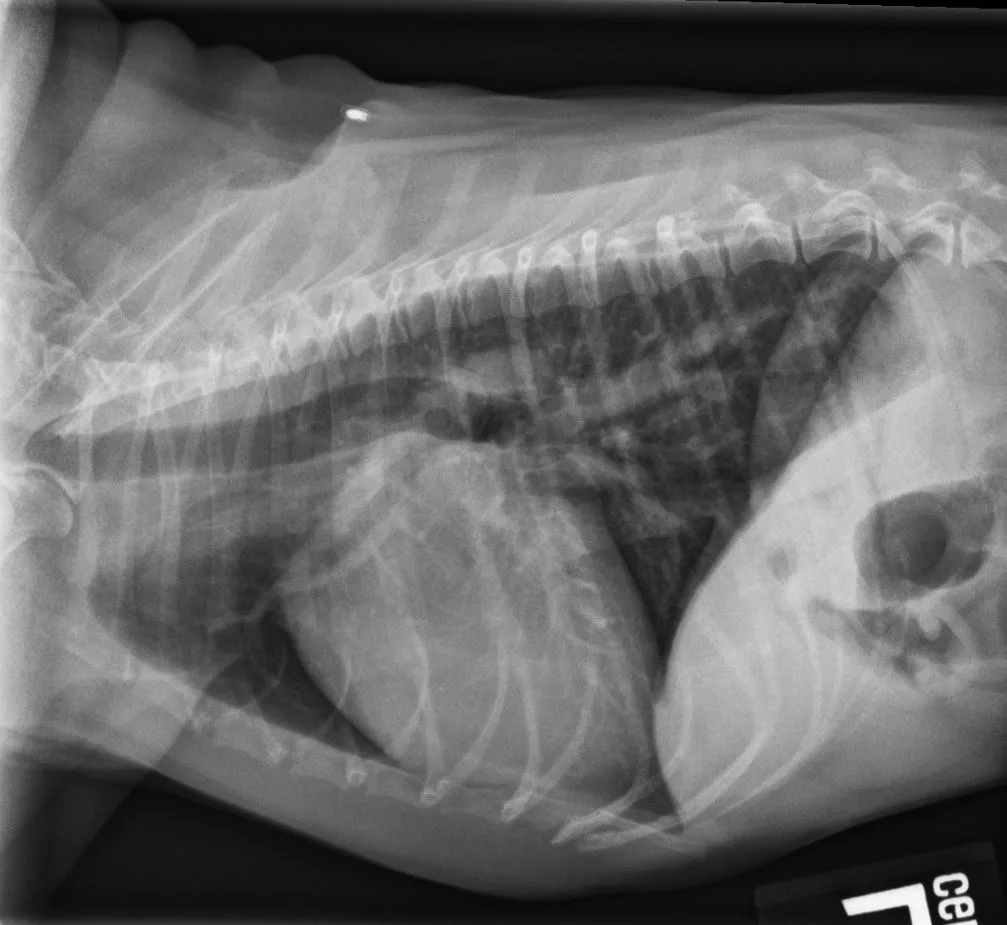
Authors of this study hypothesized that a single vertebra retains its proportion with respect to the whole body as well as the thoracic vertebral tract and using a single vertebra without shape and dimension alterations might allow objective evaluation of cardiac silhouette dimensions, even in the presence of thoracic spine alterations. The authors thus sought to create a novel method, heart:single vertebra ratio (HSVR), for radiographic evaluation of cardiac silhouette size. Length of each vertebral body from the fourth to eighth thoracic vertebrae, including respective caudal intervertebral space, was measured, as well as the cardiac long and short axes. HSVR was calculated by dividing the sum of the cardiac long and short axes by the length of each vertebral body. Radiographs of 80 dogs were included in the final assessment.
Substantial agreement was seen between VHS and HSVR determined using the seventh thoracic vertebra (HSVRT7). Results also indicated that when determining HSVRT7 is not possible, ratios using other vertebrae can be used in the following order of preference: HSVRT8, HSVRT5, and HSVRT6; however, these ratios showed slightly lower agreement with VHS compared with HSVRT7. Using the fourth thoracic vertebra (HSVRT4) was least favorable due to its lower, although still acceptable, agreement with VHS. Good to excellent inter- and intraobserver agreement for HSVR was noted.
The authors concluded that determining HSVR can overcome the intrinsic limitation of using VHS in patients with spinal alterations, including alterations that involve thoracic vertebrae between T4 and T8, representing a more objective evaluation of cardiac silhouette dimensions.
… The Takeaways
Key pearls to put into practice:
HSVR has potential value for cardiac size assessment in patients with vertebral malformations (eg, spondylosis deformans, reduced intervertebral disk spaces, hemivertebrae, butterfly vertebrae, wedge vertebrae). Another study used a similar approach, with the manubrium as an alternative.3
Assessment of normal values and breed differences for HSVR is needed before this method can be useful in clinical practice.
Regardless of the specific technique used, consistently using the same method to objectively assess cardiac size on thoracic radiographs is recommended to increase proficiency. Confidence when assessing for cardiac enlargement is more important than the method used and helps determine the appropriate time to start cardiac medications in patients with subclinical heart disease.
You are reading 2-Minute Takeaways, a research summary resource presented by Clinician’s Brief. Clinician’s Brief does not conduct primary research.