Canine Flank Alopecia
Alexander Werner Resnick, VMD, DACVD, Animal Dermatology Center
Canine flank alopecia (CFA) is a syndrome with a pathognomonic clinical appearance. Alternative terms include seasonal, cyclical, or recurrent flank alopecia.
Overview
CFA may be seasonal, with shorter durations of sunlight inducing follicular arrest (or dysplasia) and thereby lesion development. Multiple retrospective studies report that lesions most often develop between November and April. Seasonal changes in melatonin and prolactin secretion are reported in many mammals, and their presence (or lack of presence) at the hair follicles may affect hair coat growth. However, not all dogs develop lesions cyclically, nor do they all regrow hair in opposing seasons. Although lesions may appear hormone associated, they are not associated with thyroid abnormalities; however, they may be associated with follicular hormone receptors localized in the flank region.
Related Article: Canine Alopecic Dermatoses
Clinical Presentation
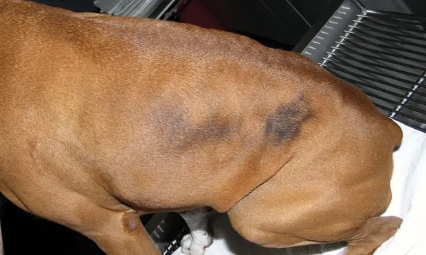
CFA lesions are initially noninflamed patches of hair coat change with irregular-to-serpiginous, well demarcated borders leading to nonscarring, noninflammatory alopecia. Exposed skin is often hyperpigmented (Figures 1 and 2). Most lesions develop in the lateral flanks in a symmetrical, but not mirror-image, pattern and extend cranially and dorsally to affect large regions; they can extend to the shoulder as well, although this is less common.
Irregularly shaped, well demarcated lesion of alopecia with hyperpigmentation in the flank region of a 2-year-old spayed boxer

Alopecic lesions similar to those in Figure 1 on a 4-year-old castrated boxer. The lesion cranial to the classical lesion appears milder.

In many patients, partial-to-complete hair regrowth may occur after several months (range, 1–14 months1; alopecia may permanently remain in individual cases). Patches of varying size may develop in subsequent years (eg, hair may change color or be patchy on regrowth). Folliculitis, an uncommon complication, can cause development of papules and crusts.
Boxers are most commonly affected; Airedale terriers, bulldogs, and schnauzers are also at increased risk. Age of onset varies, but most patients develop lesions between 3 and 6 years of age. There is no gender predilection.
Diagnosis
Because CFA presentation is sufficiently striking and location specific, diagnosis may be based on clinical appearance alone. Definitive diagnosis is determined by exclusion of more common causes of alopecia (eg, endocrinopathy, demodicosis, dermatophytosis) and biopsy of affected skin. Characteristic histologic findings include follicular atrophy, infundibular hyperkeratosis, and misshapen hair bulbs that produce a dysplastic appearance (ie, witches feet pattern).
Treatment
CFA is a cosmetic disorder typically not associated with medical abnormalities; treatment may not be necessary. Therapies that affect circadian rhythms, most notably those affected by the duration and intensity of daylight, may sometimes stimulate hair regrowth. Exposure to light boxes (used in humans for mood stabilization) has been suggested to encourage initiation of hair coat growth, although the author is not aware of published studies documenting success.
Melatonin, a major hormone affecting photoperiodic functions, has been reportedly effective for preventing hair loss and encouraging hair regrowth in CFA patients. One study reported success with SC melatonin implants or injections1; however, oral melatonin may be safer and more cost effective.
If administered, the recommended dosage is 3 mg q12h in patients weighing less than 10 kg and 6 mg q12h in patients heavier than 10 kg. Administration should be initiated each year at or just before the expected development of lesions and continue until hair regrowth is evident (~8 weeks in most cases).