Canine Dermatomyositis
Darren Berger, DVM, DACVD, Iowa State University

Dermatomyositis is an uncommon condition in dogs that predominantly affects the skin and, to a lesser extent, striated muscle.1 The exact cause is unknown, but an immune-mediated pathogenesis resulting in an ischemic dermatopathy is suspected.2 Dermatomyositis is predominantly seen in collies and Shetland sheepdogs, in which a familial predisposition is present. Other breeds may be affected, but a similar familial relationship has not been shown.
Clinical Signs
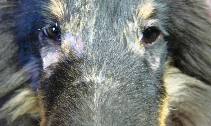
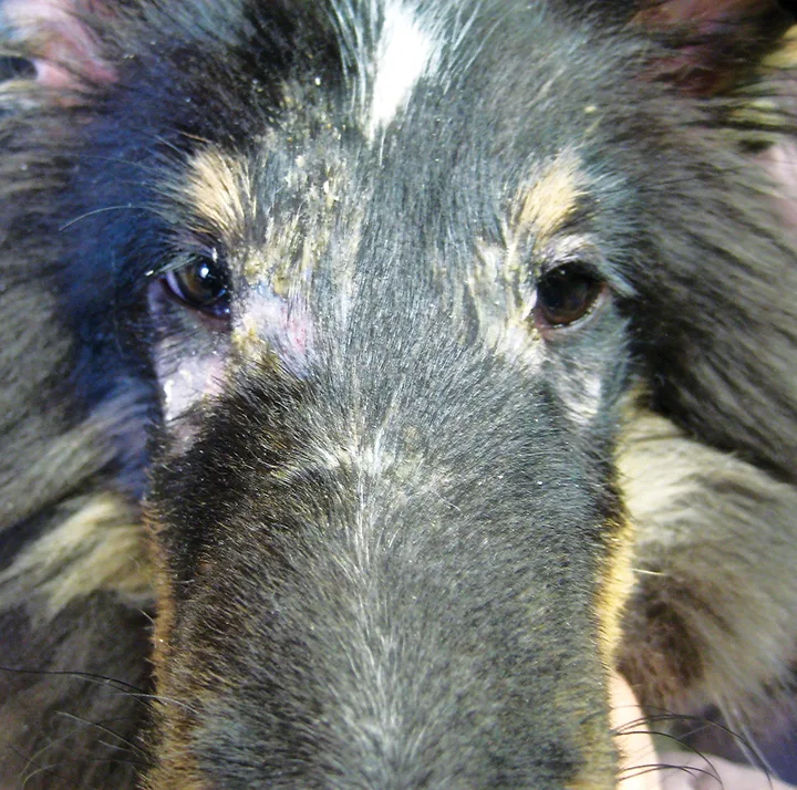
Clinical signs are variable in dogs and range from mild with spontaneous resolution to severe and debilitating. Cutaneous lesions usually appear before 6 months of age and occur on the face (periocular region, perioral region, muzzle), ears, distal extremities, and tail.2 Lesions are initially characterized by erythema, alopecia, scaling, and crusting and may progress to cicatricial alopecia. When clinical signs of myositis are present, they correlate to the severity of skin disease and manifest as difficulties with prehension of food, atrophy of masticatory and distal limb musculature, gait abnormalities, or regurgitation secondary to megaesophagus.3
Diagnosis
Diagnosis is based on signalment, recognition of typical changes, and ruling out other differentials; it is confirmed via histopathology. Muscle biopsy and electromyography aid in the diagnosis of myositis but are not routinely performed. Differential diagnoses include demodicosis, pyoderma, dermatophytosis, allergic dermatitis with secondary infections, other ischemic vasculopathies, and discoid lupus erythematosus.

FIGURE 1 A 6-month-old Shetland sheepdog with typical acute lesions of periocular alopecia, erythema, and crusting
Treatment
General recommendations include minimizing ultraviolet exposure and restricting activities that traumatize the skin, as both may exacerbate lesions. Daily supplementation with fatty acids and vitamin E (200-600 units PO q12h) may be beneficial in mild cases.3
In recurrent moderate-to-severe disease, therapy with pentoxifylline (15-25 mg/kg PO q8-12h) is commonly implemented and has historically been the drug of choice for this condition.3,4 In cases in which these recommendations do not control clinical signs, combination therapy with additional immunomodulatory agents (eg, tetracycline derivatives, niacinamide, glucocorticoids, cyclosporine, topical tacrolimus) should be pursued. Periodic flares should be expected and can be treated with short tapering courses of systemic glucocorticoids.3
Prognosis
Prognosis is highly variable and depends on disease severity as well as the ability to control signs while minimizing adverse events secondary to pharmaceutical intervention. Regardless of disease severity, affected dogs should not be bred.