Canine demodicosis is an inflammatory condition of the skin caused by increased numbers of Demodex spp mites that are normally present in low numbers in the hair follicles and sebaceous glands.1 At least 2 different species of mites (ie, Demodex canis, Demodex injai) are responsible for canine demodicosis. Demodex cornei was once considered a separate species, but literature suggests it is a variant of D canis.2
Clinical signs include patchy to diffuse alopecia, erythematous papules, pustules, and comedones. Some patients, especially those infested with D injai, may have severe seborrhea oleosa.3 Secondary infections with bacteria and/or yeast are common and may result in severe lesions (Figure 1).1

Adult-onset demodicosis with severe, methicillin-resistant deep pyoderma with hemorrhage and resulting hemorrhagic crusts
Presentation can be localized or generalized. Localized demodicosis is typically limited to <6 patchy alopecic regions on the head or limbs,1 whereas the generalized form can affect the entire body region or ≥2 feet (Figures 2 and 3).1

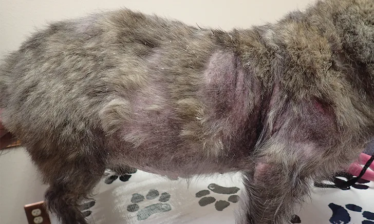
Adult-onset demodicosis with secondary Malassezia spp dermatitis and bacterial pyoderma

Closer view of patient in Figure 2; diffuse comedones can be seen
Dogs may be affected at any age. Juvenile-onset generalized demodicosis typically affects patients <1 year of age and is usually localized.1 Some cases may be due to a defect in the immune system that could be genetically linked, as evidenced by certain breed predispositions; however, immunosuppression caused by internal parasites or malnutrition may also predispose younger patients to a proliferation of Demodex spp mites.4,5 Patients that show clinical signs of adult-onset demodicosis are often >4 years of age. Most of these patients have an underlying condition—typically a disease that suppresses the immune system and causes predisposition to demodicosis.6
Demodicosis can occur in any breed, but is more frequently diagnosed in some breeds (see Frequently Diagnosed Breeds).1,4
Frequently Diagnosed Breeds15,16
American Staffordshire terrier
Boston terrier
Boxer
English bulldog
Fox terrier
French bulldog
Great Dane
Miniature pinscher
Pug
Shar-pei
Staffordshire bull terrier
West Highland white terrier
Diagnosis
Canine demodicosis is diagnosed through microscopic observation of mites in deep skin scrapings (Figure 4) and/or trichograms.1 Deep skin scrapings are obtained by using a dulled surgical blade or a spatula to scrape the skin in the direction of the haircoat until capillary oozing is achieved.The deep skin scraping or trichogram is positive when fusiform eggs, 6- or 8-legged larvae, 8-legged nymphs, or 8-legged adults are seen.1 D canis adult mites are typically 40 × 250-300 µm with a moderate-length tail. D injai adult mites are ≈40 × 350 µm with a longer tail.1 In cases in which pododemodicosis or chronic fibrosing lesions are present or the patient is a shar-pei, mites may be difficult to obtain via skin scraping, and a skin biopsy may be required for diagnosis.1

Adult D canis mite seen on a deep skin scraping (magnification, 10×)
In patients with adult-onset demodicosis, additional diagnostics (eg, CBC, serum chemistry profile, thyroid function tests, diagnostic imaging) may be required to identify underlying diseases that can suppress the immune system (eg, hypothyroidism, hyperadrenocorticism, malnutrition, neoplastic disease).7
Treatment
Localized demodicosis may resolve without treatment within 2 months.1 In breeding dogs, treatment should be withheld initially to determine whether the condition will resolve on its own or progress to generalized demodicosis. If progression occurs, the dog should not be used for breeding because the disease may be passed to subsequent generations.1
In the United States, amitraz dip is the only product labeled for treatment of demodicosis.8 Extra-label treatments (eg, isoxazolines, macrocyclic lactones [eg, avermectins, milbemycins]) are used more often than amitraz because administration is easier and more convenient.9,10 Macrocyclic lactone products should not be used in breeds susceptible to the multidrug sensitivity gene (MDR1 gene, also known as ABCB1 gene) mutation (also known as ABCB1-1delta).1,11 Examples of macrocyclic lactones include oral ivermectin or doramectin, topical moxidectin, and oral milbemycin. The isoxazoline class of parasiticides (eg, afoxolaner, fluralaner, lotilaner, sarolaner) is used more routinely due to less frequent adverse effects and increased ease of use.12-14
Table: Treatment Options for Canine Demodicosis
a The only labeled treatment is amitraz every 2 weeks. The treatment plans recommended in this table are extra label but are supported by research and the author’s personal experience.
Patients should be monitored with monthly deep skin scrapings from the 3 to 5 most severely affected areas, with treatment continuing for 30 days after the second negative monthly deep skin scraping.5 A skin scraping is considered negative when no dead or live mites are observed. A patient is not considered cured until 2 negative deep skin scrapings are obtained 1 year apart.1 Juvenile-onset generalized demodicosis has a higher chance of being cured in patients with no underlying health concerns. Patients with adult-onset demodicosis may require chronic therapy unless an underlying disease process is discovered and treated.1
Conclusion
A thorough understanding of this disease and communication with pet owners is important (see Demodicosis Talking Points). Demodex spp mites are natural inhabitants of dogs’ skin and sebaceous glands. A weakened immune system can allow mite populations to increase, resulting in alopecia, erythema, and crusting. Localized and generalized forms of canine demodicosis can occur, and onset tends to occur in patients <1 year of age (ie, juvenile-onset generalized demodicosis) or >4 years of age (ie, adult-onset demodicosis). Localized demodicosis may resolve without treatment, whereas adult-onset demodicosis is often associated with an underlying condition that requires concurrent therapy.
Demodicosis Talking Points
Following are key points to communicate to owners:
Canine juvenile-onset generalized demodicosis is more likely to be localized and respond to appropriate treatment or resolve without medical intervention.
Juvenile-onset generalized demodicosis can be hereditary, and dogs with this condition should be neutered.1
Underlying conditions should be considered when lesions occur for the first time in patients >4 years of age.
Treatment may fail if an underlying condition and/or secondary skin infection is not treated concurrently or therapy is discontinued too early.
The patient is considered cured if a negative deep skin scraping is obtained 1 year after the last negative deep skin scraping.1
Approximately 10% of patients cannot be cured.These patients may require long-term therapy