Bulla Radiography
Radiography of the tympanic bulla can help provide information on the integrity of the middle ear. For a complete clinical picture, at least 2 views should be taken. In a true lateral view, both bullae can be identified, but interpretation can be difficult since the bullae are superimposed on each other. In comparison, right and left lateral oblique views provide good visualization of each individual bulla. The rostrocaudal view provides excellent visualization of both tympanic bullae but can be technically difficult because of the need for careful positioning of the animal on its back with the endotracheal tube removed. Ventrodorsal and dorsoventral views also allow visualization of both bullae, but superimposition of the petrous temporal bones can obscure fine detail.
Lateral Oblique Views
1. Normal Canine Tympanic Bulla: Normal tympanic bulla in a dog appears as a thin-walled, crisply outlined structure with a smooth external border (arrow).
2. Normal feline tympanic bulla. Air shadow from external ear canal is superimposed on tympanic bulla.
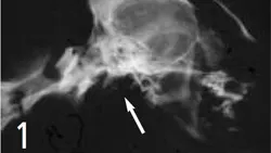
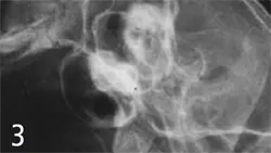
3. Magnified view of a normal feline tympanic bulla
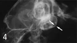
4. Feline Tympanic Bulla: Otitis Media: Bulla in a case of otitis media in a cat shows thickening of wall (arrows) and more generalized increase in opacity.
Dorsoventral View
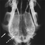
1. Air shadows in the external ear canals (arrow) help with orientation, but distortion that masks the bullae (dashed arrow) occurs because of superimposition of petrous temporal bones.
Rostrocaudal Views
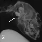
1. Feline Bullae with Inadequate Positioning: Rostrocaudal view in cats requires near-perfect positioning. Failure to open the mouth adequately (as in this case) leads to loss of skyline view of the bullae, which are superimposed on the skull (arrow).
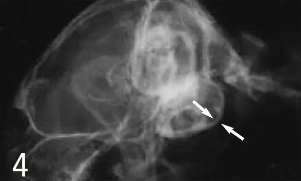
2. Feline Bulla: Otitis Media: Increased opacity of right bulla (arrow) in a cat, suggestive of otitis media
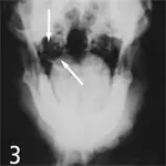
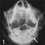
3. Canine Bulla: Normal bulla in the dog appears as thin-walled symmetrical bone opacities at the base of the skull (arrows). Overlying soft tissue can give false impression of middle-ear pathology.