Blood Transfusion Basics
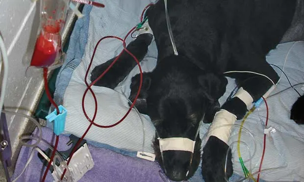
Anemia, a common condition in dogs and cats, is primarily caused by blood loss, hemolysis, or decreased erythropoiesis.
In addition to being evaluated for an underlying cause, patients with severe anemia often require red blood cell (RBC) transfusion. Packed RBCs and fresh whole blood are the two main blood products for the treatment of anemia in dogs and cats.
The choice of blood component should be tailored to the patient’s needs. Although both packed RBCs and fresh whole blood are hemoglobin sources for improving oxygen-carrying capacity, whole blood has the advantage of providing clotting factors and platelets to help with hemostasis when necessary. However, this advantage is provided at the cost of increased infusion volumes, which all patients may not tolerate.
Plasma-containing products were initially used to improve oncotic pressure in patients with hypoalbuminemia. With the widespread availability of synthetic colloids and their superior ability to improve oncotic pressure, however, there are fewer indications today for whole blood transfusions.
Blood Typing
Safe administration of blood products is important because of the potential for adverse transfusion reactions. Although a sample can be submitted to an outside laboratory for blood typing, the turnaround times are impractical. In-house blood typing kits are easy to use, provide quick results, and are fairly accurate for blood typing both species.
Crossmatching
If an appropriate blood-type unit is used, a crossmatch is not required for the first lifetime transfusion in either dogs or cats. Because antibody formation to the first transfusion may occur after 3 to 5 days, crossmatching should be performed for subsequent RBC transfusions.
Sensitization from birthing has never been proven in dogs or cats. While compatibility testing helps identify and prevent possible acute hemolytic reactions, immune reactions can occur, even with appropriately cross-matched units.
Posttransfusion Monitoring
Patients should be monitored closely for at least 60 minutes after receiving a blood transfusion. Fluid balance status should be reassessed, especially in patients with unstable cardiovascular status.
Sensitization to any RBC antigens may occur even with compatible units, and delayed transfusion reactions may take several days to manifest. Although delayed reaction (ie, hemolysis, icterus, purpura) is a well-known clinical problem in human patients, it occurs rarely in small animals.
Blood Group Systems
DogsThe blood group system recognized in dogs is the dog erythrocyte antigen (DEA) system; DEA 1.1 is the most antigenic and is used for typing purposes. A first-time RBC transfusion can be performed using DEA 1.1–negative blood without blood-typing because DEA 1.1–negative dogs are considered universal donors.
CatsIn cats, AB is the recognized blood group system. Most cats are type A, some are type B, and type AB is rarely seen; the frequency of each type varies geographically and among breeds. Blood typing is imperative in cats because they have pre-formed alloantibodies to the opposite type with no universal donors.
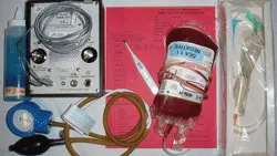
You Will Need:
Blood unit
Transfusion administration set*
Transfusion monitoring table for patient’s record
Thermometer
Blood pressure monitoring device and supplies*Most blood transfusion administration sets incorporate a 150- to 260-micron pore size filter that prevents blood clots or debris emboli. Neonatal 18-micron pore size filters exist; however, these may cause in vitro hemolysis and tend to obstruct easily, particularly with larger blood volumes (>20 mL), and are not recommended.
Step-By-Step Blood Transfusion Basics
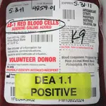
Step 1. Verify the blood product unit size, number, and expiration date as well as the donor species and blood type. Perform a visual inspection to detect any macroscopic abnormalities in color and consistency. Bacteria- contaminated blood often appears brown or purple because of deoxygenation, hemolysis, and methemoglobin formation. Also evaluate the unit for the presence of clots.
Author Insight: Warming refrigerated units is only necessary in neonates, hypothermic patients, or patients receiving large blood transfusion volumes. Warming RBCs may lead to membrane damage and hemolysis and should be avoided; blood should not exceed 37°C (98.6°F) in any instance. The unit can be brought to room temperature for 10 to 15 minutes without active warming, but for no longer, as doing so may result in bacterial growth.
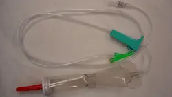
Step 2. Attach the blood transfusion administration set (A) to the blood unit, and prime it with blood (B) to eliminate all air. Then connect it to the patient’s catheter (C). Both IV and intraosseous (IO) catheters can be used. Smaller catheter sizes do not cause more hemolysis than larger ones but are the main factor in limiting the infusion rate; therefore, use a catheter with the largest available diameter.
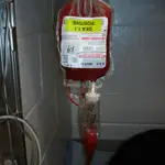
Author Insight: In neonates or when venous access cannot be achieved, IO transfusion is a good choice. About 95% of transfused blood cells will reach the systemic circulation within 10 minutes after IO administration, but the infusion rate tends to be faster and may require closer attention.
Step 3. Using a Transfusion Monitoring Chart similar to the sample shown, carefully monitor physiologic parameters and adverse reactions, including fever, hypotension, urticaria, pruritus, pigmenturia, vomiting, and shivering. Record baseline vital signs before starting the transfusion, then q15min for the first 45 minutes and q30min until the end of the transfusion. The initial infusion rate should be approximately 0.25 mL/kg for the first 30 minutes, after which the rate can be increased if no reactions are seen. The entire volume should be administered within 4 hours to prevent functional loss or bacterial growth.
Author Insight: If the infusion rate slows or stops, do one or more of the following: 1) verify patency of the infusion site; 2) examine the filter for air, excessive debris, or clots; or 3) raise the unit. If the unit is too viscous, consider adding 0.9% sodium chloride.
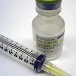
Step 4. When the transfusion is complete, flush the infusion site with 0.9% saline before initiating other infusions or drugs. Saline (0.9%) is the most compatible fluid with RBC products; hypotonic solutions cause hemolysis, and calcium-containing solutions (eg, lactated Ringer’s) can overcome the anticoagulant properties of citrate and lead to clot formation.
Author Insight: Ideally, drugs and food should be withheld during the transfusion. If a drug or fluid must be administered during the transfusion, it should be infused using a different venous access site.

Step 5. Check packed cell volume (PCV) and total solids 1 to 6 hours after transfusion. If there is no ongoing loss or hemolysis, 70% of the transfused RBCs are expected to remain in circulation. Thereafter, if fresh whole blood was used, the RBCs should have a normal lifespan (approximately 110 days in dogs, 70 days in cats). The lifespan of packed RBCs depends on the age of the unit: the longer the unit has been stored, the shorter the lifespan.
Author Insight: If a reading is obtained too soon after the transfusion, fluid compartment equilibrium may be incomplete, leading to inaccurately high PCV readings. Fluid compartment equilibrium typically occurs 30 to 60 minutes after infusion.
ALEXANDRE PROULX, DVM, is currently completing a residency in small animal emergency and critical care at University of Pennsylvania School of Veterinary Medicine. His areas of interest include acid-base disturbances and extracorporeal life support. Dr. Proulx graduated from University of Montreal School of Veterinary Medicine, after which he completed a rotating small animal internship at University of Pennsylvania Matthew J. Ryan Veterinary Hospital.
LORI S. WADDELL, DVM, DACVECC, is an adjunct assistant professor in the intensive care unit at University of Pennsylvania Matthew J. Ryan Veterinary Hospital. Her research interests include colloid osmotic pressure and coagulation in critically ill patients. Dr. Waddell is a graduate of Cornell University School of Veterinary Medicine and completed an internship at Angell Memorial Animal Hospital in Boston. After her internship, she worked as an emergency clinician in private practice and completed a residency in emergency medicine and critical care at University of Pennsylvania School of Veterinary Medicine.