Bilateral Iatrogenic Mandibular Fracture
Gottfried Morgenegg, DVM, Tierzahnarzt, Obfelden, Switzerland
Ana Nemec, DVM, PhD, DAVDC, DEVDC, University of Ljubljana, Ljubljana, Slovenia
Brook A. Niemiec, DVM, DAVDC, DEVDC, FAVD, Veterinary Dental Specialties & Oral Surgery, San Diego, California
THE CASE
A 12-year-old, 7.9-lb (3.6-kg) neutered male miniature pinscher was emergently presented to a veterinary dental specialist following a dental cleaning at his primary veterinarian. During the dental cleaning, significant periodontal disease bilaterally involving the mandibular first molars had been noted on periodontal probing. Although dental radiography and equipment to section teeth were not available at the primary veterinarian’s clinic, an attempt was made to extract the mobile teeth fully intact. However, both mandibles fractured at the level of the first molars, partially due to bone loss associated with severe periodontitis, and the patient was subsequently referred to the dental specialist.
Emergency Presentation
On presentation to the specialist, the patient had a ventrally deviated mandible due to bilateral fractures. Physical examination findings were otherwise unremarkable; all vital parameters, aside from a BCS of 4/5, were within normal range. The tongue was slightly hanging out of the mouth on the left, and slight drooling was noted from the commissures of the mouth. Oral examination was not possible in the conscious patient due to behavior.
Preoperative blood work was in reference range. An IV catheter was placed, the patient was premedicated with hydromorphone, and maropitant was administered. Anesthesia was induced with diazepam and propofol. The patient was intubated and maintained with sevoflurane (2%) and oxygen. Monitoring included body temperature, ECG, pulse oximetry, noninvasive blood pressure, and capnography. A balanced IV crystalloid solution was also administered.
Diagnostics
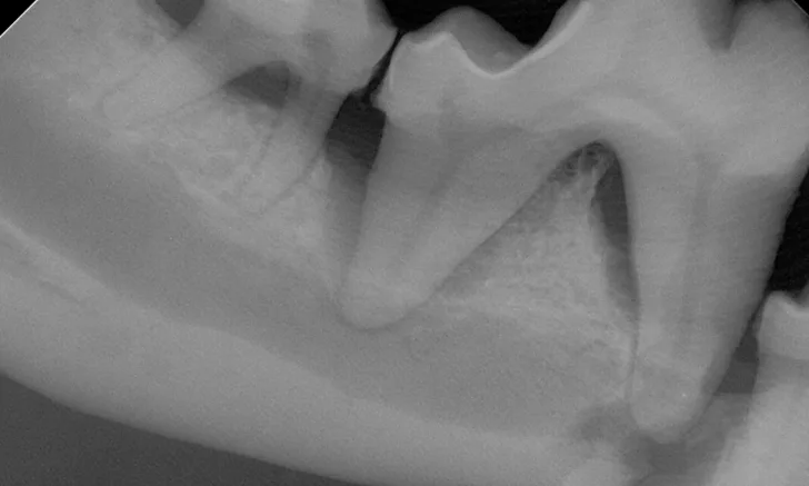
A complete oral examination confirmed stage 4 periodontal disease of numerous teeth (ie, first through fourth maxillary premolars, all maxillary molars, both maxillary canines, all remaining incisors). Dental radiographs confirmed bilateral mandibular fractures at the mesial root of the right mandibular first molar and distal root of the left mandibular first molar (Figures 1 and 2).

FIGURE 1A
Preoperative intraoral dental radiographs of the right (A and B) and left (C) mandibular first molars. There is marked alveolar bone loss secondary to periodontal disease (blue lines), as well as pathologic, iatrogenic mandibular fractures (arrows).
Treatment & Follow-Up
A regional anesthetic agent (0.5% bupivacaine [0.2 mL]) was injected where the mandibular nerve enters the mandibular canal (inferior alveolar nerve block). The affected teeth were surgically extracted to minimize distraction of the bones, the area was gently debrided, and each mandible was stabilized with a single intraosseous wire; the oral soft tissues were sutured in a simple interrupted pattern with 5–0 poliglecaprone-25 to completely cover the bone (Figure 3). This provided sufficient reduction of the fractures and adequate stability.
The patient recovered uneventfully and was discharged the same day with medication for pain management (ie, meloxicam, buprenorphine) and instructions for the owner to administer only soft foods.
The patient was presented 7 weeks later for a recheck oral examination under general anesthesia (performed in the same manner as previously). Dental radiographs demonstrated healing of the fractures (Figure 4). The interfragmentary wires were removed via an intraoral approach, and postoperative intraoral radiographs were obtained to confirm adequate healing (Figure 5). The patient recovered uneventfully.

FIGURE 3A
Postoperative intraoral dental radiographs of the right (A) and left (B) mandible following reduction and fixation with a single intraosseous wire on each side.
Discussion
Pathologic jaw fracture is a significant local consequence of chronic periodontal disease.1,2 These fractures typically occur in the mandible due to chronic periodontal tissue loss, which weakens the bone in affected areas.3 Although these fractures can occur in any area of the mandible, they are especially common near the canine and first molar teeth.4 Pathologic jaw fractures are more common in small-breed dogs as compared with large-breed dogs due to their mandibular first molars being larger in proportion to the mandible itself.5 Small-breed dogs also have a minimal amount of bone apical to the tooth root, putting this area at high risk for fracture when apical bone loss occurs.6
Pathologic jaw fractures have a guarded prognosis due to the lack of remaining bone and poor bone quality, presence of infection, low oxygen tension in the area of the fracture, and difficulty in rigid fixation of the caudal mandible.4,6 Regardless of the method of fixation used, diseased root(s) must be extracted to facilitate healing.3,7
Pathologic jaw fractures typically occur as a result of mild trauma but can also occur during dental extraction procedures (ie, iatrogenic fracture).4 Clinical awareness can help reduce risk for iatrogenic fractures during at-risk dental procedures (eg, extraction of the mandibular canines in dogs and cats, the mandibular first molars in small-breed dogs, the mandibular fourth premolars in small-breed brachycephalic animals, and teeth in any area weakened by infection or neoplasia).6
Dental radiography is critical to the proper care of dental patients, as radiography can help identify risk factors for jaw fracture (eg, alveolar bone loss).7 In cases in which severe alveolar bone loss is noted, particularly if the mandibular canine or first molar is affected, owners should be informed of the possibility of iatrogenic jaw fracture prior to extraction of the offending tooth.3,4
Regardless of the degree of bone loss, diseased teeth with minimal remaining bone can be successfully extracted using proper technique.
Multirooted teeth should always be sectioned prior to extraction. This is important because roots of most multirooted teeth are divergent, and thus root tips will break if extractions are attempted in one piece.3,7-11 Root fracture can occur even if a tooth is relatively mobile. In addition, buccal bone removal may be performed if indicated, particularly if one of the roots or part of the root has significant periodontal attachment.8
This patient was successfully managed with a single interfragmentary wire on each side. This technique was elected because it was possible to achieve very good anatomic reduction of the fractures and provide clinically acceptable stability. However, when intraosseous wires are employed to fix fractures of the body of the mandible, it is important to ensure neutralization of bending forces with tension-band wire along the alveolar margin of the mandible and avoid tooth roots. A second area of fixation can also be considered at the ventral mandibular margin with a stabilization wire, which neutralizes rotational and shear forces. This will allow proper biomechanics for healing and prevent movement during the healing period.11
Conclusion
Pet owners should be counseled about the importance of proper dental care to avoid the significant effects of periodontal disease. Further, dental radiography should always be performed prior to any extraction; it is particularly important to radiograph the mandible in small-breed dogs. Proper extraction techniques, including sectioning of multirooted teeth, should always be performed. Educating pet owners of the possibility of fracture is important from a legal aspect. In addition, patients with minimal apical bone should be referred to a veterinary dental specialist when possible.
ASK YOURSELF …
Bilateral Iatrogenic Mandibular Fracture Quiz
Take this quiz by answering the following multiple choice questions.