Bacterial Pneumonia

Profile
Definition
Systems
Bacterial pneumonia is a lung infection caused by gram-positive or -negative, aerobic or anaerobic bacteria.
Mixed infections are common.
Mycoplasma spp may be involved.
Bacterial pneumonia caused by atypical organisms such as Mycobacterium spp are not considered in this review.
Incidence
The incidence is not known.
Geographic Distribution
Distribution is worldwide.
Related Article: Infectious Respiratory Disease in a Dog
Signalment
Species
Diagnosis is much more common in dogs than in cats.
Breed Predilection
No primary breed predilection exists, although underlying disease may have a breed association.
Age
Puppies and kittens are more likely than adults to have bacterial pneumonia from primary infection or underlying viral or congenital causes (eg, cleft palate, megaesophagus, ciliary dyskinesia).
Older patients, particularly well-vaccinated dogs without exposure to organisms associated with canine infectious respiratory disease complex (CIRDC), often have underlying disease.
Causes
Primary respiratory tract pathogens include Bordetella bronchiseptica and Streptococcus equi subsp zooepidemicus in dogs and cats and Mycoplasma cynos in dogs.
Other Mycoplasma spp may be primary pathogens in dogs and cats.
B bronchiseptica has been isolated from tracheal wash fluid in 50%-70% of puppies with community-acquired pneumonia.1,2
Opportunistic infections are typically caused by organisms residing in the oropharynx or nasopharynx.
Organisms associated with CIRDC can cause primary bacterial pneumonia or can predispose the animal to opportunistic infections.
A recent study found canine parainfluenza virus in 7 of 20 dogs diagnosed with bacterial pneumonia involving organisms other than B bronchiseptica.3
Related Article: Megaesophagus
Risk Factors
Primary respiratory infections are acquired from exposure to other infected animals or fomites.
Exposure could occur in locations such as shelters or rescue housing, pet stores, breeding operations, boarding or grooming facilities, and dog parks.
Exposure to animals (or the toys or bowls of animals) with unknown vaccination history or incomplete vaccinations against respiratory pathogens increases potential for infection.
Animals with immature or compromised immune systems and those that have not been vaccinated against respiratory pathogens are at increased risk if exposed to infected animals or materials.
Numerous underlying conditions or predisposing factors lead to bacterial pneumonia from opportunistic organisms (see Underlying Conditions & Predisposing Factors).
Pathophysiology
Bacteria typically enter the lungs through the airways, either through inhalation of primary infectious agents or aspiration of oral, pharyngeal, esophageal, or gastric contents.
Hematologic seeding of the lung with bacteria from elsewhere in the body can also occur.
This route of infection is likely under-diagnosed because thoracic radiographs typically show a diffuse interstitial to alveolar pattern rather than the more classic gravity-dependent alveolar pattern of bronchogenic or aspiration pneumonia.
A hematogenous origin was documented in more than half of cats with bacterial pneumonia based on necropsy.4
Clinical signs result from damage to the airway epithelium, influx of inflammatory cells, edema, and mucus hypersecretion.
History
Signs are most often acute (ie, of only a few days’ duration) but can be present for several months.
Cough is a common sign.
Signs of compromised lung function include (in order of increasing severity): increased resting respiratory rate, increased respiratory effort during mild activity, respiratory distress, and cyanosis.
Systemic inflammatory signs (eg, lethargy, inappetance) may be present.
Careful questioning is indicated to identify risk factors, predisposing conditions (eg, anesthesia), and exposure to primary pathogens.
Physical Examination
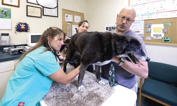
Auscultation of crackles, loudest in the cranioventral lung lobes, is the classic finding.
Auscultation must include positioning the stethoscope in the axilla, under which the cranial lung lobes are positioned.
Although crackles in any lobe in a previously healthy animal is an important finding, absence of crackles does not eliminate possibility of pneumonia.
Crackles were reported in only 9% to 25% of dogs with aspiration pneumonia.5,6
Increased respiratory effort, orthopnea, respiratory distress, or cyanosis is present with sufficient compromise.
Signs of systemic inflammation, such as fever and depression, may be present.
Fever is present in only about one-third of dogs with aspiration pneumonia5,6 and is considered uncommon in cats with lower respiratory tract infections.7
A normal temperature does not rule out bacterial pneumonia.
Related Article: Respiratory Distress: Diagnosis & Treatment at a Glance
Diagnosis
Definitive Diagnosis
Confirmation of diagnosis requires cytologic demonstration of septic inflammation in an airway specimen—usually a tracheal wash (Figure 1).
A positive bacterial culture confirms the involvement of specific organism(s) and provides valuable information for guiding antibiotic selection.
Previously healthy patients with mild-to-moderate disease and no risk factors for antibiotic-resistant infection may not require airway sampling and are usually treated empirically based on other clinical information.
Severely compromised animals may be too unstable to tolerate collection of an airway specimen and are likewise treated empirically without delay.
Tracheal wash is most likely to provide a representative specimen in pneumonia of airway origin.
Bronchoalveolar lavage (BAL) may be indicated if radiographs suggest interstitial disease, as this is more likely than tracheal wash to provide a representative specimen in such cases.
If possible, tracheal wash (or BAL) should be performed before antibiotic administration.
After antibiotic administration, cytology and culture are often falsely negative for infection.*
Infection with multiple species of organisms is common.
Mycoplasma spp infections are difficult to diagnose because organisms are not apparent cytologically, and a specific culture must be requested.
Polymerase chain reaction (PCR) testing is available and is considered more sensitive than culture.
The role of Mycoplasma spp as a primary pathogen is unknown, so coinfections should be considered.
The value of Mycoplasma spp testing in individual cases is debatable, but the author recommends testing in dogs with suspected exposure to CIRDC organisms and in cats.
A presumptive diagnosis of pneumonia is made with suggestive history, physical examination, CBC, and/or thoracic radiographic findings.

Tracheal wash fluid from a dog with septic inflammation. At low magnification (A), neutrophils and mucus are abundant. At high magnification (B), intracellular and extracellular rod-shaped bacteria can be seen. (Modified Wright’s stain)
Differential Diagnosis
The main differential diagnosis for patients with airway origin pneumonia is sterile aspiration pneumonia.
In dogs and cats, however, bacterial infection is common following aspiration.
An important differential is congestive heart failure.
Cats with congestive heart failure can have patchy, asymmetric alveolar infiltrates on thoracic radiographs that mimic those of bacterial pneumonia.
Attention should be paid to the size of the heart and pulmonary veins.
Echocardiography and ProBNP can be useful.
Diagnostic differentials for hematogenous pneumonias are many and include infection with other organisms (eg, viruses, protozoa, fungi), interstitial lung disease (including eosinophilic bronchopneumopathy), neoplasia, hemorrhage, and pulmonary edema (among others).
Dogs with typical CIRDC have self-limiting tracheobronchitis.
Bacterial pneumonia can occur as a result of bacteria associated with CIRDC or secondary to viral infections of CIRDC.
Cats with bacterial pneumonia can present with signs consistent with feline asthma.
Laboratory Findings
Neutrophilic leukocytosis with left shift supports a diagnosis of bacterial pneumonia.
Only 40% to 60% of dogs with aspiration pneumonia have increased numbers of bands5,6; therefore, bacterial pneumonia cannot be ruled out on the basis of a normal CBC.
Imaging
Airway-origin pneumonia most often results in a bronchoalveolar pattern, most severe in one or more of the gravity-dependent lung lobes (right or left cranial and right middle lobes).
Hematogenous origin pneumonia most often results in a diffuse interstitial or mixed pattern, which may be most severe in the caudal–dorsal lung lobes.
Bacterial infection associated with a foreign body may result in a localized interstitial, alveolar, or consolidated pattern.
Thoracic radiographs should be scrutinized for signs of concurrent or predisposing disease within the lung or in other organs, particularly megaesophagus, bronchiectasis, lung lobe torsion, or cavitary disease.
Other Diagnostics
Ultrasound can be performed in areas of lung consolidation if neoplasia, foreign body, abscess, or lung lobe torsion are differentials.
Computed tomography is often pursued in patients with recurrent or nonresponsive pneumonia to search for underlying or complicating disease (eg, foreign bodies, bronchiectasis, neoplasia, abscess).
Bronchoscopy is likewise often conducted in patients with recurrent or nonresponsive pneumonia.
Bronchoscopy is performed if airway specimens are needed but tracheal wash is unrewarding or radiographic location of disease (eg, primarily interstitial) makes a successful tracheal wash less likely.
Postmortem Findings
If death occurs before antibiotic administration, septic inflammation with isolation of organisms from tissue culture or PCR is expected.
Characteristic findings may be obscured by prior treatment.
In the absence of a known cause for pneumonia prior to death, virus isolation or PCR should be considered and all other organs carefully investigated.
Treatment
Inpatient or Outpatient
Hospitalization is required for oxygen administration in distressed patients, for fluid administration in dehydrated patients, and for IV antibiotic treatment for patients with systemic inflammatory response syndrome or with esophageal or GI disease that would interfere with the absorption of oral medications.
Stable, hydrated patients can be treated as outpatients.
Patients with suspected or confirmed infection with organisms associated with CIRDC or with multidrug-resistant bacteria should be managed using isolation protocols.
Medical
Antibiotic administration and maintenance of systemic hydration are indicated for all patients.
Nebulization of sterile saline may improve mucociliary clearance in patients with severe clinical signs and lung consolidation.
Antibiotics can be administered by nebulization, but this route is reserved for resistant organisms to avoid systemic toxicity of parenteral drugs and is considered inadequate as the sole antibiotic coverage for patients with pneumonia.
Bronchoconstriction may be induced by nebulization with medication other than sterile saline.
Systemic antibiotics are the primary treatment for bacterial pneumonia.
Coupage may also help to promote airway clearance and can be performed for 5-10 minutes several times daily.
Coupage is indicated following nebulization.
Encouraging activity and frequent turning of recumbent patients is likely more effective, however, and coupage should not replace these treatments.
Nutritional Aspects
Attention to nutrition is indicated for all patients with infection, particularly if systemic signs are present.
Gastric tubes or parenteral nutrition may be considered for patients with esophageal or GI disease.
Activity
Clearance of exudate from the lung is enhanced by exercise as a result of changes in body position, increased tidal volumes, and stimulation of productive cough.
Patients should be encouraged to exercise to the extent their respiratory function allows.
Patients that are unable to stand should be rotated from side to side and to sternal recumbency every 2 to 4 hours.
Client Education
Specific discussion points will depend on disease severity, the origin of the bacterial pneumonia, and underlying or predisposing problems.
Pneumonia secondary to CIRDC warrants a discussion of the benefits and limitations of routine vaccination as well as isolation of the patient to avoid spread of disease.
Alternative Therapy
Shorter courses of antibiotics are used in humans with community-acquired pneumonia, which may be analogous to dogs that have bacterial pneumonia as a complication of CIRDC.
In the author’s experience, short courses of antibiotics in dogs or cats with chronic or recurrent disease, or with underlying or predisposing factors, are not likely to be successful.
Medications
Drugs and Fluids
Ideally, antibiotic selection is based on results of bacterial culture and susceptibility testing (Table).
Pending culture results, or if testing was not performed, some general guidelines follow.
Monitoring for response to treatment is indicated regardless of how antibiotics are selected, with a positive response expected within 72 hours.
For patients with mild or moderate disease, consider amoxicillin with clavulanate, cephalexin, or trimethoprim–sulfonamide.
If pneumonia is suspected to be associated with CIRDC, consider doxycycline for its historic activity against many Bordetella spp isolates and efficacy against Mycoplasma spp.
This drug has a narrow spectrum for gram-negative organisms and should not be used as the sole drug for patients with moderate-to-severe signs.
For patients with severe or life-threatening disease, broader- spectrum antibiotic coverage may be prudent and might include ampicillin/sulbactam and a fluoroquinolone, ampicillin/sulbactam and an aminoglycoside, or clindamycin and a fluoroquinolone.
It is generally recommended that antibiotics be continued for 1-2 weeks beyond the resolution of clinical and radiographic signs of disease.
Treatment for 4-6 weeks is typical.
Shorter courses may be sufficient in acute cases with no ongoing underlying or predisposing conditions, such as those with pneumonia associated with CIRDC.
Careful attention to hydration status, with administration of IV fluids as needed, is essential to maximize mucociliary clearance and support local circulation.
Over-hydration may contribute to pulmonary edema.
Bronchodilators (theophyllines or b-agonists) may be indicated in cats with associated bronchoconstriction.
They might be helpful in distressed dogs through potential mechanisms such as improved mucociliary clearance or anti-inflammatory effects, but they can potentially worsen ventilation–perfusion mismatch and should be used cautiously.
Corticosteroids at anti-inflammatory doses may be indicated in life-threatening situations.
Whether they are ultimately beneficial or harmful remains undetermined.
Table. Antibiotics for Bacterial Pneumonia
Contraindications
Diuretics, such as furosemide, contribute to dehydration of airway secretions and should be avoided.
Cough suppressants are generally not indicated and could interfere with coughing’s beneficial effects.
Dogs with severe cough, such as that resulting from concurrent or underlying tracheal collapse, chronic bronchitis, or CIRDC, may benefit from judicious suppression to minimize fatigue and break a cycle of cough followed by inflammation.
Precautions & Drug Interactions
Fluoroquinolones can delay the clearance of theophylline; if both are chosen, the dosing interval of theophylline should be increased 2-fold.
Follow-up
Patient Monitoring
Patient monitoring is critical.
Selected antibiotics may not be sufficient to treat all organisms involved, and unidentified or untreatable underlying conditions may delay recovery or result in re-infection.
A positive response to antibiotics is expected within 72 hours of initiating treatment.
Patients with moderate-to-severe disease requiring hospitalization are monitored by careful physical examination at least twice daily.
Thoracic radiographs and CBC are performed every 48-72 hours in most cases.
Additional monitoring may be indicated depending on predisposing, concurrent, or complicating conditions.
Following discharge, patients are evaluated every 1-2 weeks until signs have resolved.
Depending on abnormalities detected initially, monitoring typically includes CBC and thoracic radiography along with a complete physical examination.
It may be sufficient in patients with mild disease to follow up with periodic phone calls and a single office visit to allow for a complete physical examination.
Patients with persistent or recurrent signs may require more aggressive follow-up with airway specimen collection or more advanced diagnostic testing, such as CT or bronchoscopy.
Complications
Acute complications include sepsis, respiratory failure, and death.
Chronic complications are rare but include lung abscess formation.
Failure to respond to treatment or recurrence suggests either an ongoing underlying or predisposing problem and/or infection with a resistant organism.
In General
Relative Cost
$$ to $$$$$
Bacterial pneumonia in otherwise healthy individuals resulting from a single event, such as aspiration following anesthesia or as a complication of CIRDC, may resolve readily and completely with a course of antibiotic therapy on an outpatient basis.
Patients with severe disease may require intensive care for 1 week or longer.
Patients with underlying problems that cannot be corrected are candidates for recurrent episodes.
A study of dogs with aspiration pneumonia reported a median hospitalization of 3 days (range, 0-11 days) at a mean cost of $2581 (range, $241-$10400).5
Prognosis
Short-term prognosis can be good to grave.
Long-term prognosis depends on the origin of infection and underlying or predisposing factors.
Two studies of dogs with aspiration pneumonia reported 72% and 77% survival, respectively.5,8
Prevention
Risk for contracting primary bacterial pneumonia or pneumonia as a complication of CIRDC can be minimized by avoiding contact with susceptible populations and by routine vaccination, as indicated by individual patient circumstance.
Careful attention to technique can minimize risk for aspiration from iatrogenic causes.
Aggressively seeking and managing predisposing factors is indicated in patients with unexplained disease.
*Based on anecdotal clinical experience.
BAL = bronchoalveolar lavage, CIRDC = canine infectious respiratory disease complex, PCR = polymerase chain reaction
ELEANOR C. HAWKINS, DVM, DACVIM, is assistant department head for the small animal internal medicine and emergency/critical care service at North Carolina State University. Her clinical, research, and teaching interest is in respiratory disease. Author of the respiratory section of the Small Animal Internal Medicine textbook, Dr. Hawkins has been section editor of Current Veterinary Therapy and The 5-Minute Veterinary Consult. She has served as board chair of the American College of Veterinary Internal Medicine.