Anuric Renal & Hepatic Failure in a Dog
Alyssa Sullivant, DVM, MS, DACVIM, Mississippi State University
Todd Archer, DVM, MS, DACVIM (SAIM), Mississippi State University
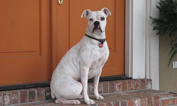
Betsy, a 5-year-old spayed boxer, was presented for a 2-day history of vomiting, anorexia, and azotemia.
History & Examination
Betsy appeared dull and depressed but responsive. Mucous membranes were hyperemic, and capillary refill time was <1 second. Clinical dehydration was not detected. BCS was 5/9 with mild abdominal distension. Mild hypothermia (98.6F [37C]) was present, but heart and respiratory rates were normal. Severe generalized abdominal pain and ptyalism were noted.
Betsy was up-to-date on core vaccinations. More than a year before presentation, leptospirosis vaccination with serovars Canicola, Icterohaemorrhagiae, Pomona, and Grippotyphosa had been administered. Betsy received monthly heartworm and topical flea preventives but no other medications. She had access to several acres of wooded property and had suffered superficial skin wounds from a raccoon a week before presentation. Although a rabies booster was indicated, it was not administered. Betsy was hospitalized and treated with IV fluids and maropitant (1 mg/kg SC once a day), as well as enrofloxacin (5 mg/kg IV once a day) and ampicillin (20 mg/kg IM 3 times a day) for presumptive infection, but no improvement was seen. Abdominal distention developed, and Betsy was referred for specialty care. The referring veterinarian observed that Betsy did not urinate overnight despite receiving fluid therapy.
Diagnostic Results
CBC was unremarkable, but serum chemistry profile identified several abnormalities (Table 1). Urinalysis revealed trace glucosuria, 1+ proteinuria, and a urine specific gravity of 1.026. A urine culture yielded no bacterial growth. Baseline ammonia was significantly elevated. Severe hypertension was present (240 mm Hg systolic Doppler) despite administration of hydromorphone. Fundic examination was normal, and no evidence of uveitis was noted. Thoracic radiography revealed a diffuse, unstructured intersitial pattern, generalized mild bronchial pattern, and mild pleural effusion.
SIGNIFICANT LABORATORY RESULTS
Abdominal ultrasonography revealed a small urinary bladder, bilateral hyperechoic renal cortices, ascites, and a hyperechoic pancreas.
Abdominal ultrasonography revealed a small urinary bladder, bilateral hyperechoic renal cortices, ascites, and a hyperechoic pancreas. A SNAP (idexx.com) cPL (canine pancreas-specific lipase) was abnormal. Abdominocentesis yielded a pure transudate with fluid potassium (4.16 mEq/L) and creatinine (6.0 mg/dL) values equal to serum chemistry values. A urinary catheter was placed, but no urine was obtained despite administration of 5 L of IV fluids over the previous 24 hours. Leptospirosis titers were submitted.
Diagnosis
Acute renal failure and liver failure due to presumptive leptospirosis
Related Article: Acute Renal Failure
Treatment & Outcome
Betsy was hospitalized for 14 days with IV fluid therapy to match intake and output and to resolve azotemia. Fluid overload, anuria, and hypertension resolved 6 hours after the start of treatment. A furosemide constant rate infusion (CRI; 1 mg/kg/hr) was necessary for the first 24 hours to maintain normal urine output. Treatment for leptospirosis was initiated with ampicillin–sulbactam, although ampicillin alone is the recommended leptospirosis treatment. IV antibiotics were continued until the patient ate well, at which time doxycycline (5 mg/kg PO twice a day) was administered for 3 weeks. Supportive care was also provided (Table 2). Betsy was discharged after 14 days of hospitalization with oral doxycycline, S-adenosylmethionine (SAMe) + silybin (20 mg/kg PO once a day), famotidine (0.5 mg/kg PO twice a day), and aluminum hydroxide (100 mg/kg PO divided daily with meals). Mild azotemia and mild hyperphosphatemia persisted at discharge. Initial leptospirosis titers were Leptospira interrogans serovar Grippotyphosa (1:100), L interrogans serovar Bratislava (1:400), and L interrogans_serovar Autumnalis (1:100). Three weeks later, convalescent titers revealed an 8-fold rise in _L interrogans serovar Grippotyphosa titers (1:800). Recheck bloodwork at 1 and 3 months was normal.
Related Article: Leptospirosis in Dogs
Treatment at a Glance
Antioxidant
Caution: May exacerbate a vitamin K-dependent coagulopathy, especially with high doses of vitamin E
| | Vitamin K (1 mg/kg SC twice a day)* | Coagulopathy associated with liver failure |
*Treatment administered in-hospital only
Discussion
Leptospirosis is a worldwide zoonotic disease.1 Acute renal failure with or without acute hepatic failure commonly develops. Pancreatitis, disseminated intravascular coagulation, hepatic failure (without renal involvement), and pulmonary hemorrhage syndrome have also been documented.1
Related Article: Quiz: Leptospirosis
Leptospirosis is seen most commonly in late fall and after periods of heavy rainfall.2 Organisms are shed in the urine of reservoir hosts (eg, raccoons, rodents) and transmitted through contact of mucous membranes or broken skin with contaminated water or soilwhere the organism can survive for extended periodsor directly through ingestion of infected tissues.2 The incubation period is 1 week, and leptospiremia is present for 3 to 4 days before the organisms reach the renal tubules, where they are shed in the urine.2 Doxycycline is the treatment of choice and should be initiated as soon as oral therapy is tolerated to resolve urine shedding and decrease the risk to humans.2 Penicillins are effective at clearing leptospiremia and can be used until doxycycline is tolerated; however, they do not clear the carrier state.2 Treatment with doxycycline is recommended for 2 to 3 weeks, and prognosis can be fair at 50% to 80% survival.2 Complete renal recovery may take 3 to 4 weeks, and residual renal damage may persist.2
Related Article: Leptospirosis: Protecting Your Patients & Staff
BCS = body condition score, CBC = complete blood count, CRI = constant rate infusion, MAT = microscopic agglutination test, PCR = polymerase chain reaction, SAMe = S-adenosylmethionine