Anal Sacculectomy
OverviewAnal sac disease in dogs and cats is thought to be due to thick secretions; a change in anal muscle tone or consistency of stool; bacterial infection; small, underdeveloped ducts; or a combination of these factors. Aerobes and anaerobes are pathogens that are usually associated with any infection involving the anal sacs; anaerobes are probably responsible for the foul odor sometimes associated with anal sac infection.
Most dogs affected with anal sac disease are toy breeds, although an occasional large- or giant-breed dog may be affected. Cats have anal sac disease on rare occasions. Tenesmus; dyschezia; scooting, biting, and/or licking at the perineum; and tail chasing can occur alone or in various combinations. Medical therapy often fails to completely resolve the problem. Anal sacculectomy is indicated for chronic anal impaction, chronic sacculitis, abscesses, and as part of the treatment for perianal fistulas.
What You Will Need
Saline or antiseptic solution
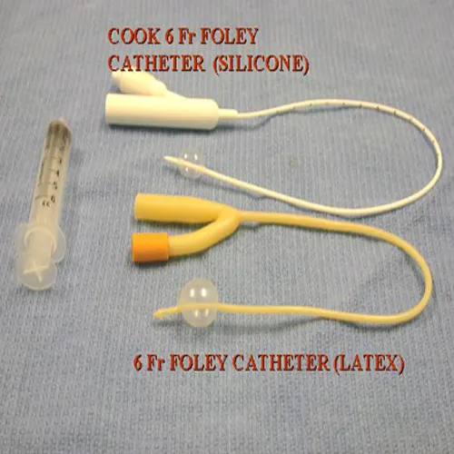
6-French silicone or latex catheter
Saline-soaked gauze
Foley catheter
Surgical instruments
3-0 or 4-0 multifilament suture
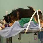
1. Place the animal in sternal recumbency and tie the tail cranially over the back.
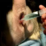
2. Express contents of the anal sacs and flush gently with saline or an antiseptic solution.
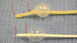
3. Place a 6-French silicone or latex catheter into the anal sac. In most smaller dogs and cats, the silicone catheter fits into the sac better without backing out due to the shorter distance between the tip of the catheter and the distal extent of the inflated cuff (arrows).
4. Place the tip of the catheter gently into the anal sac. Pack the rectum with a sufficient amount of saline-soaked gauze to provide a barrier against fecal soilage.
PROCEDURE PEARLSofter suture material is preferred to avoid irritation to the tender perianal area.
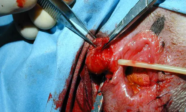
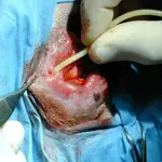
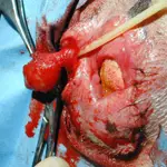
5. Gently fill the bulb of the Foley catheter with saline until the anal sac can be easily palpated below the skin. In most cases, 1 to 1.5 ml of saline is adequate. Occasionally, the inflated bulb will back out of the sac as it is distended. Place a suture across the anal duct opening to prevent this. Once distended, the bulb makes identification and palpation of the gland simple. The protruding catheter allows the surgeon or the assistant to place gentle traction on the sac during the dissection. Use scissors to dissect down to the wall of the anal sac, which can be easily identified by its grayish color compared with the reddish color of the external anal sphincter. Place slight traction on the catheter as dissection proceeds and the sac is removed.
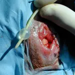
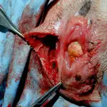
6. After gently incising the skin overlying the anal sac, use sharp and blunt dissection to isolate and expose the anal sac. Be sure to stay close to the inflated sac to minimize injury to the external anal sphincter muscle fibers.
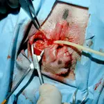
7. Once the sac is dissected free, excise the duct along its length toward the duct opening.
8. Flush the pocket that remains after removal of sac vigorously with saline. Repeat flushing three or four times and appose the skin.
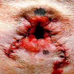
9. I prefer to use 3-0 or 4-0 absorbable multifilament synthetic suture to appose the skin because it is softer than nylon and less irritating to the tender perianal region. Sutures are removed in 2 weeks.
PROCEDURE PEARLPain management during postoperative care is critical.
The owner should be counseled thoroughly about postoperative care. Significant discomfort and irritation may be present for the first 3 to 5 days. Pain management is critical. In my experience, most animals require an Elizabethan collar to prevent self-mutilation.