Abdominal Abscess in a Dog
As part of an international shelter dog rescue and relocation program, Rufus, a 15-month-old intact male, mixed-breed dog from Chaguanas, Trinidad, was flown with several other dogs to Florida, where he was adopted by a family in Tampa.
Rufus had free-range access to a fenced backyard, where he enjoyed chasing frogs.
Examination
About 4 months after adoption, Rufus presented with a swollen area adjacent to the penis. The lesion was red, warm to the touch, and associated with an ecchymotic spot on the skin. Other than an elevated body temperature of 105°F (normal, 100.5°F–102.5°F), there were no other significant clinical findings.
Diagnostics
CBC and serum biochemistry profile were declined by the owner, but the urinalysis results were normal.
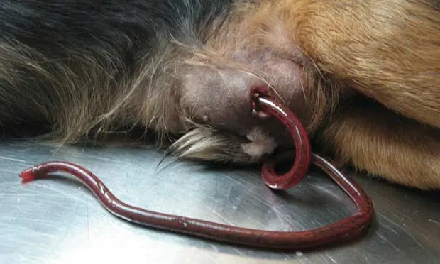
Exploratory Surgery
A peripheral IV catheter was placed and Rufus was sedated. Substantial purulent exudate was drained from the abscess, and after copious flushing with normal saline and hydrogen peroxide, the end of a bright red worm was visible. Further sedation, careful manipulation, and continued probing facilitated extraction of a 60-cm long dark red worm (Figure 1 above, Worm extracted from the abscess while the dog was under sedation).
Ask Yourself...
What is the likely way that Rufus became infected?
Where else in the Americas might one stumble on infection with this parasite?
What additional diagnostics would be advisable and why?
Diagnosis
Dioctophyma renale infection
The worm in this case was the unique but recognizable bright red Dioctophyma renale. The sex of the recovered worm was not determined by the attending veterinarian; however, its size (60 cm) suggests an immature female worm, as adult females can reach 1 meter long while males are typically 30 to 40 cm long.
View life cycle of Dioctophyma renale.
Fortunately, this was the only worm present in this dog. When both male and female worms are present in the abdominal cavity, they typically mate and produce massive numbers of eggs, causing granulation tissue to form throughout the abdominal mesentery.
The typical site of maturation of D renale worms is the kidney, often the right kidney, of the dog or other host. When the kidney is involved, the worm basically erodes away almost the entire cortex and medulla, leaving only the capsule. The kidney can contain one or more female and male worms. When both sexes are present in the kidney, eggs may pass in the urine of the infected dog.
The number of adult worms found in dogs has ranged from 1 to 34 worms, with as many as 14 worms in the right kidney.
Treatment
Treatment typically involves surgical extraction. This case was fairly atypical because of the worm exiting the body through a cutaneous lesion, although similar cases have been reported in other animals and even humans.
Outcome
Rufus was discharged with an appropriate course of cephalexin and returned to the clinic for recheck examination 1 week after worm extraction. Abdominal ultrasonographs revealed that both kidneys were normal with no signs of additional worms in the peritoneal cavity.
Did You Answer...
The life cycle of D renale involves a small freshwater oligochaete annelid as the first intermediate host; the ingested eggs develop into larvae that in turn are ingested by freshwater fish or frogs (paratenic hosts). Dogs and other mammals (definitive hosts) then become infected when they consume the frog or fish (Figure 2, View life cycle of Dioctophyma renale). Because Rufus was known to chase frogs in his backyard, it is assumed that he captured a frog and consumed a portion of it.
In the Americas, most cases of D renale infection have been reported in Canada, northern United States, and Brazil. In addition, this parasite is found in Eurasia, and the typical hosts are considered to be mink and other mustelids.
When D renale infection involves the kidneys, ultrasonography will confirm erosion of the cortex and medulla, along with the presence of worms. In this case, the ultrasound was performed to determine if perhaps any additional worms were present, although none were detected.
DWIGHT D. BOWMAN, MS, PhD, is president of the Companion Animal Parasite Council. He has been on the faculty of Cornell University as a parasitologist in the College of Veterinary Medicine since 1987. Dr. Bowman’s work concentrates on the practical control of parasites in domestic animals and the study of parasite biology as it relates to control. His research interests include soil-transmitted parasites and pathogens, wildlife parasites, and visceral larva migrans. Dr. Bowman earned his degrees at Tulane University.
MIGUELLA MARK-CAREW, BS, is a comparative biomedical sciences PhD candidate at Cornell University, where her research involves understanding potential epidemics at the population level, with specific attention to Giardia duodenalis infection both in the United States and abroad. She recently received a Fulbright grant to study in Trinidad and Tobago, where she worked with Dr. Kevern Sawh. Ms. Mark-Carew completed undergraduate studies at Dartmouth College.
KEVERN SAWH, DVM, is head veterinarian at Animedics Pet Hospital in Chaguanas, Trinidad. He is actively involved in the Trinidad and Tobago Veterinary Association and provides veterinary assistance to local shelters and animal welfare nongovernmental organizations in Trinidad. Dr. Sawh received his DVM from University of West Indies School of Veterinary Medicine.