In the Literature
Tully M, Milley C, Bourgeois A, Bidot W. Comparison of cytological findings between direct impression smears and adhesive slides in canine superficial bacterial folliculitis. Vet Dermatol. 2024. doi:10.1111/vde.13279
The Research …
Skin cytology is vital for diagnostic investigation of dermatologic disease. Cytology can help determine presence of microorganisms (ie, bacteria, yeast, other fungi) and type and severity of inflammation based on presence of neutrophils, eosinophils, and other WBCs.1 Techniques include direct impressions, acetate tape impressions, and direct impressions with adhesive slides.2 Although cytology is routinely recommended, the optimal technique for detecting certain microorganisms or inflammatory cells is unclear.
This study compared direct impression cytology with plain glass slides to direct impression cytology with adhesive slides. Fifty superficial bacterial folliculitis lesions (ie, epidermal collarettes, crusts, papules) were sampled from 15 client-owned dogs. Each slide was evaluated by 3 clinicians for polymorphonuclear cells, keratinocytes, nuclear streaming, intra- and extracellular debris, and miscellaneous findings, which notably included Malassezia spp yeasts.
Plain glass slides yielded significantly more extracellular cocci and nuclear streaming than adhesive slides, and adhesive slides yielded more keratinocytes and miscellaneous debris. The techniques were equivalent for intracellular cocci, intra- and extracellular rods, and polymorphonuclear cells. Because the samples were obtained from lesions of diagnosed superficial bacterial folliculitis, the authors concluded that use of plain glass slides is more appropriate when bacteria detection is the goal, and use of adhesive slides may be more appropriate when keratinocyte detection is the goal.
… The Takeaways
Key pearls to put into practice:
Skin diagnostics can be a source of frustration. If impression cytologies with plain glass slides (Figure) or acetate tape are not producing diagnostic results, incorporation of adhesive slides in routine dermatologic examinations should be considered. For example, acetate tape is better suited for achieving adequate contact with interdigital spaces or claw folds compared with glass slides, but examination of samples obtained with both a plain glass slide and either an adhesive slide or acetate tape from other body regions may be useful because acetate tape and adhesive slides may yield more miscellaneous debris.
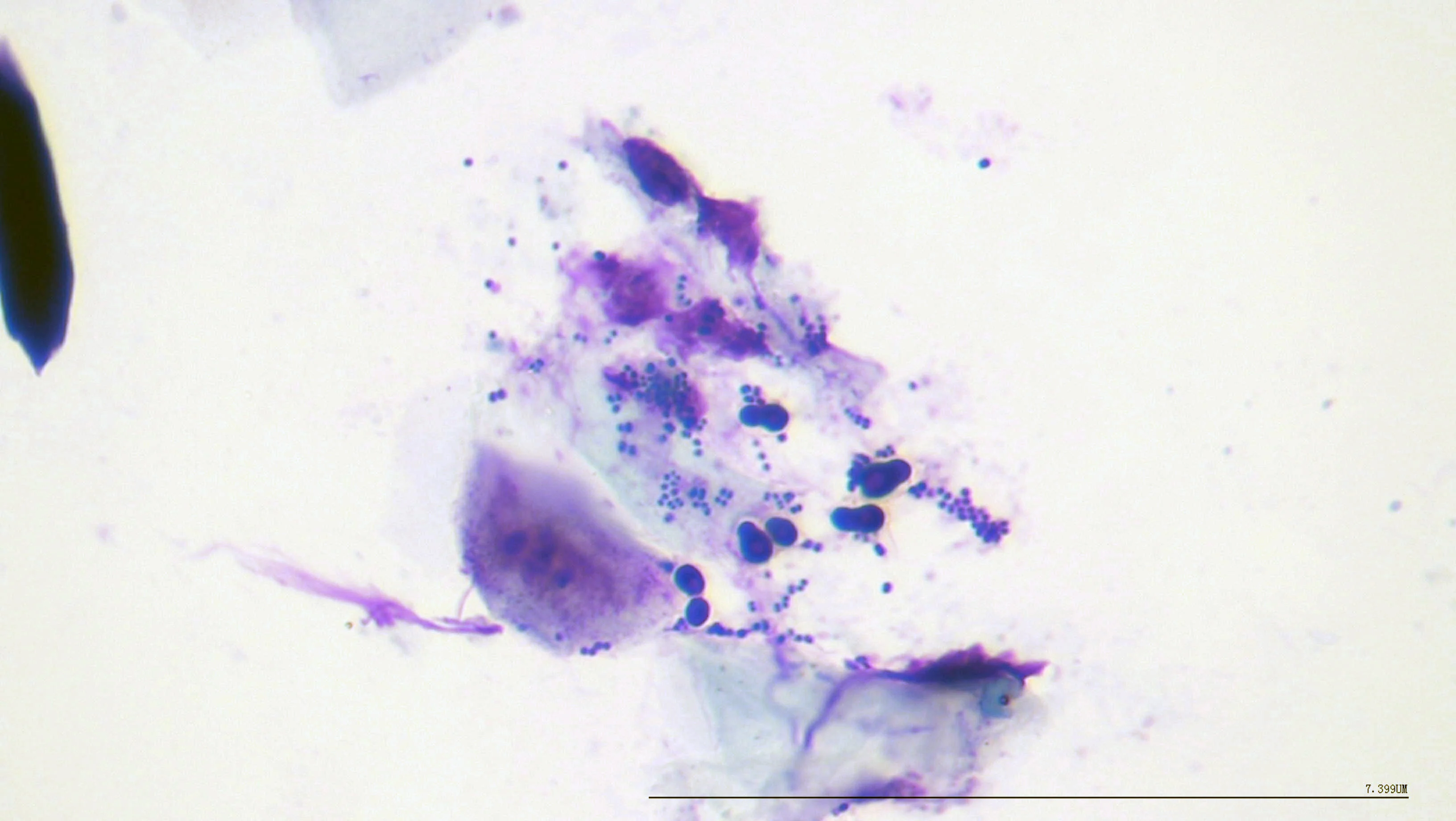
Plain glass impression cytology from superficial pyoderma and Malassezia spp dermatitis in a dog showing coccoid bacteria, Malassezia spp yeast, nuclear streaming, and keratinocytes. 1,000× magnification
Although performing multiple cytology techniques takes additional time, accurate detection of microorganisms and inflammatory cells can lead to better treatment outcomes; use of all 3 techniques on different lesions and body regions can also help develop a preferred technique. Technicians with appropriate training can read cytology slides quickly and enter the results into the medical record, thus improving clinic workflow.
Antibiotics are frequently administered for treatment of skin disease3; however, antibiotic use should be critically evaluated in consideration of antimicrobial stewardship. Cytologies are a rapid and low-cost method to determine whether and to what degree bacteria are present, which can inform clinical decisions.
You are reading 2-Minute Takeaways, a research summary resource presented by Clinician’s Brief. Clinician’s Brief does not conduct primary research.