Treatment of Acute Hemoabdomen in a Dog

Penelope, a 6-year-old, 44-lb, spayed pit bull terrier, presented 10 minutes after being struck by a vehicle traveling 35 miles per hour.
Physical Examination
Penelope had a heart rate of 200 bpm, respiratory rate of 32 breaths/min, and temperature of 99.8°F. She was obtunded and laterally recumbent. Mucous membranes were pale pink to white and moist with a prolonged capillary refill time (CRT) of 3 seconds. Femoral pulses were weak. She had normal heart and lung sounds. Penelope’s abdomen was tense and painful on palpation. No orthopedic or neurologic abnormalities were detected. A Doppler blood pressure of 80 mm Hg was recorded.
Laboratory Results
PCV was 40% (range, 35%–55%) and total protein (TP) was 3.2 g/dL (range, 5.2–7.8 g/dL). Irregularities in the biochemistry profile were present (See Table). Coagulation times (prothrombin and partial thromboplastin) were normal. The lactate level was 6.4 mmol/L (range, 1.2–4.14 mmol/L).
Related Article: Placement of an Abdominal Wrap to Control Hemodynamics
Treatment
IV fluid resuscitation was initiated with several rapid infusions totaling 70 mL/kg of crystalloids (Plasmalyte-A), 20 mL/kg colloids (hydroxyethyl starch), and the only unit of stored whole blood (450 mL). Penelope was administered 0.1 mg/kg of IV hydromorphone but remained tachycardic (180 bpm) and had weak pulses. An abdominal fluid wave was noted; abdominocentesis revealed a hemorrhagic effusion with a PCV of 34% and TP of 2.8 g/dL. A recheck revealed peripheral blood PCV of 20% and TP of 2.2 g/dL.
Table: Abnormal Biochemistry Profile Findings
CRT = capillary refill time, TP = total protein
ASK YOURSELF...
Benefits, Risks, & Techniques for Autologous Transfusions
Hemorrhage into the thoracic or abdominal cavities can be quickly collected and administered IV. Autologous blood has been demonstrated to be lifesaving, safe, and advantageous (See Suggested Reading below).
Benefits
Simple to collect and administer
Frequently sterile
Contains functionally superior cells compared with banked blood
Does not require warming
Decreased risk for transfusion reactions from foreign protein
Can be less expensive than banked blood
Readily available
Risks
Potential complications attributable to autotransfusion include:
Hypocoagulation
Hemolysis
Sepsis
Dissemination of malignancy
Embolism
Techniques
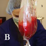
Blood can be suctioned out of the abdominal or thoracic cavity with centesis or direct suction (in a surgical case). Blood should be collected directly into a sterile suction canister, iv fluid bag (Figure 1), commercial blood bag, or syringe (in smaller patients). Passing the blood through a filter is recommended. Adding anticoagulant is often unnecessary unless hemorrhage is ongoing at collection. Monitoring PCv, Ts, electrolytes, and renal values is recommended.
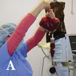
Autologous blood was collected in a sterile fashion and transferred into an empty sterile IV bag. It was administered IV through a filter, as shown here.
Autologous blood was collected in a sterile fashion and transferred into an empty sterile IV bag. It was administered IV through a filter, as shown here.
CRT = capillary refill time, TP = total protein, TS = total solids
Treatment Options
Mild hemorrhage often responds to IV replacement with crystalloids and colloids. Rapid infusions (over 10–15 min) of 20 to 30 mL/kg of crystalloids and 5 mL/kg of colloids are necessary and may need to be repeated. Patients with coagulopathies or severe hemorrhage may require blood products. When blood banks are stressed or large volumes are required, autologous transfusion remains a viable option.
The risk:benefit ratio weighs heavily in favor of autotransfusion for the resuscitation of select patients, even despite neoplasia or gross contamination, when facing exsanguination and death.
Related Article: Current Thoughts on Coagulopathy Testing

Outcome
Penelope was taken to surgery where a large splenic laceration was identified as the source of ongoing hemorrhage; 900 mL of autologous blood was collected intraoperatively with a Poole suction tip into a sterile canister, transferred to a sterile IV bag, and administered IV. A splenectomy was performed, and the remainder of the exploratory laparotomy was unremarkable. Penelope recovered uneventfully from anesthesia and was discharged 48 hours later. Four weeks later, she was doing well and had returned to normal activity.
The Take-Home
Life-threatening hypoperfusion (shock) can be easily detected during examination by assessing heart rate, gum color, CRT, mentation, and pulse quality.
A drop in TS relative to PCV is an early pathologic sign of hemorrhage; decreases in PCV may not occur until fluids have been administered.
Imaging should not delay treatment for shock.
To provide intravascular volume and oxygen carrying capacity, autologous blood transfusion can be easily performed in patients suffering from life-threatening cavitary hemorrhage when blood resources are unavailable or exhausted.