Treating Tooth Resorption in Dogs
Jan Bellows, DVM, FAVD, DAVDC, DABVP, All Pets Dental, Weston, Florida
This is the second installment of a two-part presentation investigating tooth resorption in dogs. While the first installment (January 2013 issue) outlined the various classifications of tooth resorption, the following segment maps treatment options and techniques.
Intraoral radiograph of the rostral mandibles revealing advanced external resorption affecting both canine teeth
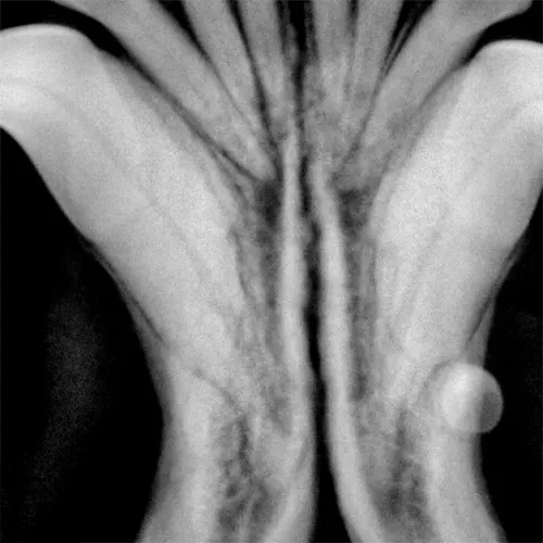
Appropriate therapy for tooth resorption will depend on clinical and radiographic appearance, as well as owner preference once treatment options have been discussed. Intraoral radiography is central to the decision-making process; if intraoral radiography is unavailable and tooth resorption is clinically apparent, the patient should be referred. If referral is not an option, the tooth should be extracted to eliminate oral bacterial invasion. Without intraoral radiography, root fragments may be left behind during extraction, potentially causing painful problems later.
1. Recheck
When resorption begins deep in the alveolus on the root surface, the periodontal ligament and root parts may be replaced with bone- or cementum-like tissue via a noninflammatory process. If the lesion does not appear to extend into the oral cavity, it can be monitored for progression q6mo. Watchful waiting is indicated in cases with minimal root involvement not extending into the oral cavity; follow-up intraoral radiography q6–12mo is recommended accordingly. If the resorption area approaches or extends into the oral cavity, extraction is the treatment of choice.
Related Article: Treating Tooth Resorption in Dogs
Related Article: Feline Tooth Resorption—Extraction of Retained Premolar Root
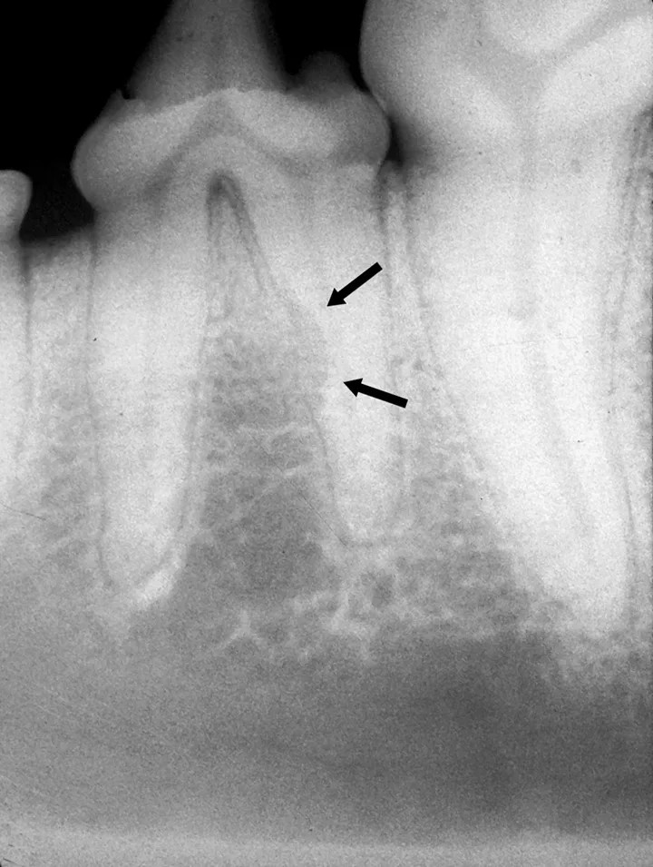
FIGURE 1A
Intraoral radiograph of the left mandibular fourth premolar; the distal root is affected by external resorption that has not extended into the oral cavity.
2. Reduce
In rare forms of tooth resorption, the root’s dental hard tissue is replaced by bone. The root will appear to have less radiopacity when compared with adjacent normal roots. The remaining crown’s height can be reduced to a subgingival level via amputation and sutured closed.
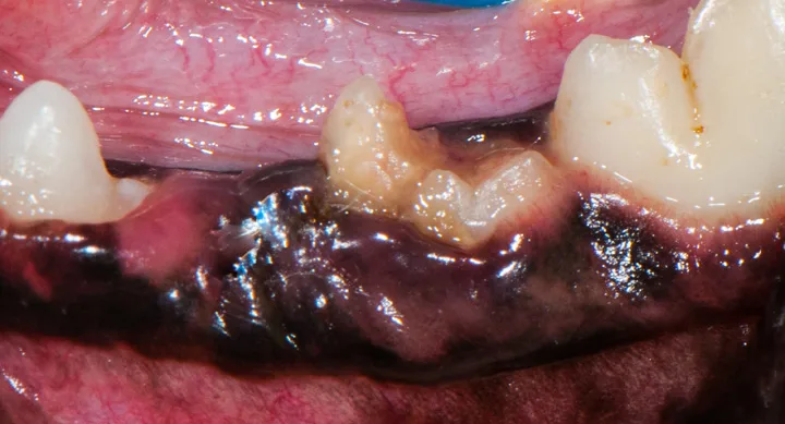
FIGURE 1A
Resorbing deciduous (primary) left mandibular fourth premolar.
3. Root Canal Therapy
Root canal therapy, an advanced procedure with guarded prognosis, can be used to treat cases of internal and external tooth resorption.1 Because tooth resorption in dogs is idiopathic and possibly progressive, long-term success of root canal therapy may not affect the clinical course of tooth destruction.
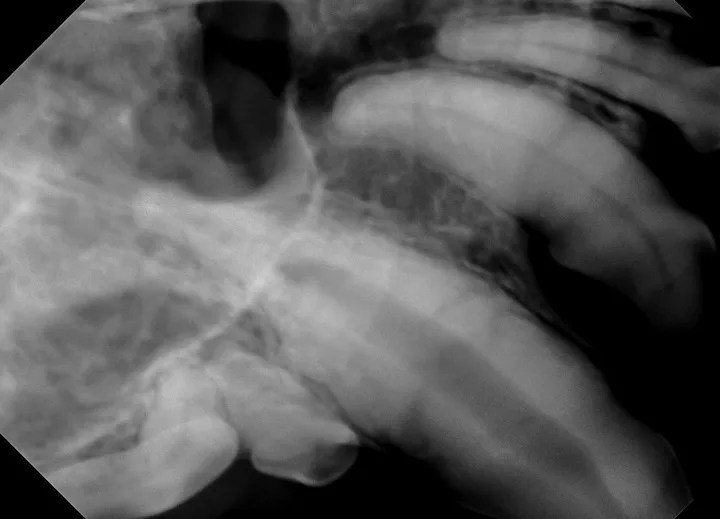
FIGURE 1A
Resorption of the right maxillary canine root in a 3-year-old English bulldog.
4. Remove
When resorption begins near the cementoenamel junction and progresses toward the crown, dentin and enamel loss near the gingival attachment often exposes the sensitive dentinal tubules and pulp to the oral environment. Subsequent inflammation of surrounding tissues can lead to increased sensitivity. Once the resorption extends into the oral cavity, surgical extraction via flap exposure is indicated.
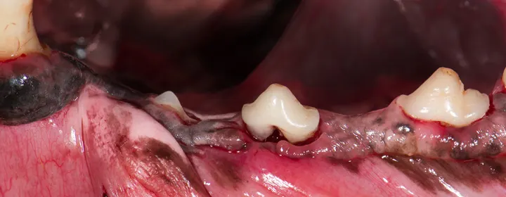
FIGURE 1A
Left mandibular cheek teeth.
5. Refer
Removing all dental hard tissue is often difficult in cases of canine tooth resorption. Extraction of an affected tooth (or multiple teeth) is technique sensitive and can be time intensive. Referral is often the best option (avdc.org can help locate a local veterinary dentist).