Subcutaneous Hemorrhage

A 10-year-old, neutered female mixed-breed dog was presented with a history of swelling and bruising of the shoulder and thorax.
History. One month ago, the referring veterinarian aspirated blood from the swelling and treated the dog with vitamin K. When examined this week, the patient was listless, had labored respiration, and was reluctant to walk, favoring a swollen right forelimb. Her packed cell volume was 13%. The dog was referred to the University of Florida Veterinary Medical Center for evaluation.
Physical Examination. Massive subcutaneous hemorrhage was present in the right axillary region. The mucous membranes were pale and respiratory and heart rates were rapid. The rectal temperature was normal.
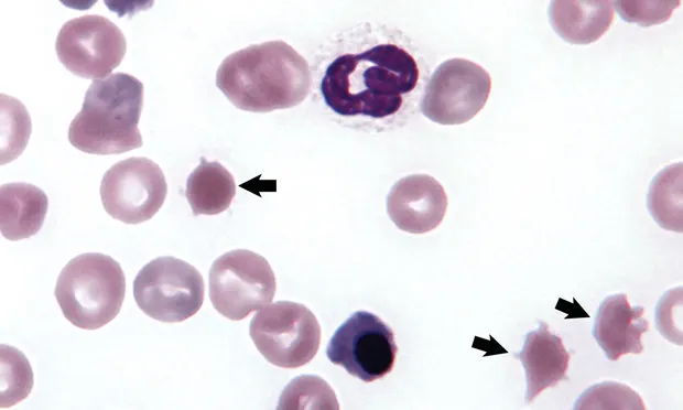
Laboratory Analysis (see Table). The dog had macrocytic, normochromic anemia with absolute reticulocytosis. The platelet count was low, the total plasma protein was slightly decreased, and the plasma was slightly yellow. Erythrocyte morphologic abnormalities included 2+ anisocytosis, 2+ polychromasia, and 2+ poikilocytosis. The prothombin time, the activated partial prothombin time, and the fibrin degradation products were all normal. The differential leukocyte count revealed leukocytosis with mature neutrophilia and monocytosis. Large numbers of nucleated erythrocytes were present. The predominant erythrocyte shape abnormality is shown in Figure 1, above.
Figure 1. A neutrophil, two nucleated erythrocytes (metarubricytes), four polychromatophilic erythrocytes, and three abnormally shaped erythrocytes (arrows) in a blood film (Wright- Giemsa stain; original magnification, ´1000).
ASK YOURSELF...
What specific term is used to describe erythrocytes with the shape abnormality shown by the arrows in Figure 1?
What disorders have been associated with this shape abnormality in dogs?
What additional tests might you considerperforming?
Diagnosis: Hemangiosarcoma with acanthocytosis
The regenerative anemia and slightly decreased total plasma protein concentration may be attributed to the ongoing hemorrhage present in this dog, but the presence of hyperbilirubinemia (slightly yellow plasma) and acanthocytes suggests that some component of erythrocyte destruction might also be present. The mild thrombocytopenia present was not sufficient to account for the persistent hemorrhage observed and no coagulation abnormalities were identified to explain the bleeding. Acanthocytes are spiculated erythrocytes with irregularly spaced, variably sized spicules (Figure 2, blue arrows). Acanthocytes have been recognized in animals with liver disease.1 They have also been reported in dogs with disorders that result in erythrocyte fragmentation such as hemangiosarcoma, disseminated intravascular coagulation, and glomerulonephritis.1-3 Lesser numbers of schistocytes (fragmented erythrocytes) were also present in blood from this dog (Figure 2, orange arrows).
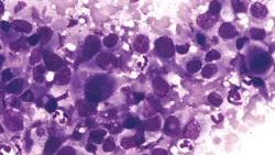
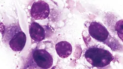
Additional Test Results. Radiographs of the thorax revealed pleural thickening and pleural effusion. Radiographs of the abdomen were unremarkable. About 100 ml of blood was drained from the chest cavity by thoracentesis. Tissue was collected surgically from the right brachial plexus area. An anaplastic tumor involving spindle-shaped cells (sarcoma) was diagnosed from the stained impression smears (Figures 3 and 4). Given the history, physical findings, and laboratory findings, a diagnosis of hemangiosarcoma was made. The client elected euthanasia and granted permission for a cosmetic necropsy. A large thoracic mass was identified and removed for histopathologic examination. Histopathologic examination of the subcutaneous tissue and the thoracic mass confirmed the diagnosis of hemangiosarcoma.

Figure 3. Tissue imprint from surgical biopsy (Wright-Giemsa stain; original magnification, ´3000).

Figure 4. Tissue imprint from surgical biopsy (Wright-Giemsa stain; original magnification, ´ 1300).
Hemangiosarcoma. Hemangiosarcomas are malignant tumors of vascular endothelial origin. Tumor rupture and hemorrhage are common and sudden death can occur when the rupture occurs in a critical area or from acute bleeding into a body cavity.4 A reticulocytosis in response to blood loss anemia is generally expected within a few days after hemorrhage has occurred. Nucleated erythrocytes are often observed in association with reticulocytosis and are sometimes more numerous than generally seen in regenerative anemia.3 Microangiopathic erythrocyte destruction may also occur within the tumors, and a high incidence of acanthocytes has been reported in dogs with hemangiosarcoma and fragmentation anemia.<sup3, 5sup> The presence of schistocytes in the blood of this dog indicates that some degree of erythrocyte fragmentation occurred.
Acanthocyte formation is primarily associated with hemangiosarcomas involving the liver in dogs with radiation-induced tumors.5 The association between liver tumors and acanthocytes has not been identified in dogs with naturally acquired hemangiosarcomas.2 Thrombocytopenia is common in dogs with hemangiosarcoma. Mild to moderate thrombocytopenia may occur following massive blood loss.3,6 Thrombocytopenia may also result from consumption of platelets within the tumor (localized thrombosis) or in association with disseminated intravascular coagulation, which sometimes occurs in dogs with hemangiosarcoma.6,7 Hemostatic test results did not support the occurrence of disseminated intravascular coagulation in this dog, but localized thrombosis within the hemangiosarcoma may have resulted in platelet consumption and erythrocyte fragmentation.5 As seen in this case, a leukocytosis with neutrophilia is common in dogs with hemangiosarcoma.<sup3, 4sup>
DID YOU ANSWER...
Acanthocytes
Liver disease and disorders that result in erythrocyte fragmentation, such as hemangiosarcoma, disseminated intravascular coagulation, and glomerulonephritis
Biopsy the axillary region and perform diagnostic imaging of thorax and abdomen
SUBCUTANEOUS HEMORRHAGE • John W. Harvey
References
Blood and bone marrow of domestic animals. Harvey JW. Atlas of Veterinary Hematology-Philadelphia: WB Saunders, 2001, pp 29-30.2. A retrospective study of canine hemangio-sarcoma and its association with acantho-cytosis. Hirsch VM, Jacobsen J, Mills JH. Can Vet J 22:152-155, 1981.3. Clinical and haematological features of haemangiosarcoma in dogs. Ng CY, Mills JN. Austral Vet J 62:1-4, 1985.
Hemangiosarcoma. In MacEwen EG, Withrow SJ (eds): Small Animal Clinical Oncology-Philadelphia: WB Saunders, 2001, pp 639-646.
Microangiopathic hemolytic anemia associated with radiation-induced hemangiosarcomas. Rebar AH, Hahn FF, Halliwell WH, et al. Vet Pathol 17:443-454, 1980.
Hemostatic abnormalities in dogs with hemangiosarcoma. Hammer AS, Couto CG, Swardson C, Getzy D. J Vet Intern Med 5:11-14, 1991.7. Localized consumptive coagulopathy associated with cutaneous hemangiosarcoma in a dog. Rishniw M, Lewis DC. JAAHA 30:261-264, 1994.