Step-by-Step: The Neurologic Examination
Cheryl L. Chrisman, DVM, MS, EdS, DACVIM (Neurology)
The neurologic examination is a series of observations and tests done to answer the following four questions:
Is a lesion in the nervous system present?
Where is the lesion located (focal or multifocal)?
How severe is the lesion?
Is the disease worsening, improving, or staying the same? (serial neurologic examinations)
Step-by-Step: How to Perform a Neurologic Examination
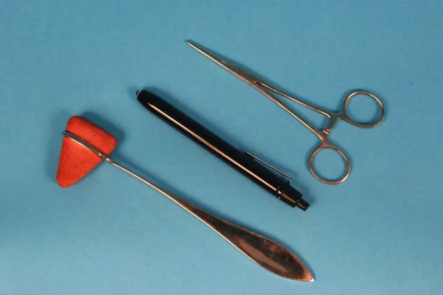
What You Will Need
Penlight
Percussion hammer
Pair of hemostatic forceps
Initial Observations & Cranial Nerves
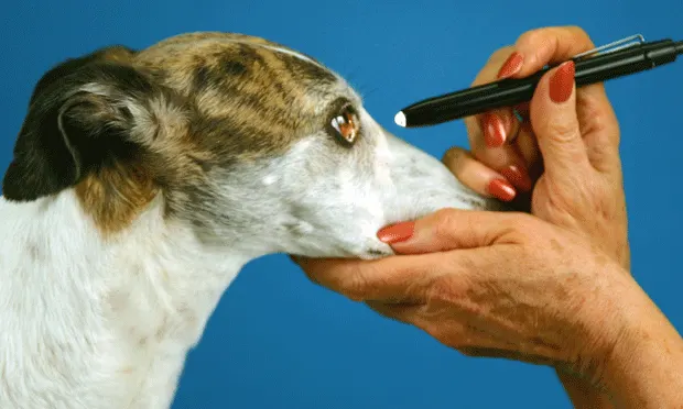
1. Initial observations
Mentation, head posture, and coordination and function of some cranial nerves can be directly observed. The animal will sniff and eat if the olfactory nerves (CN1) are functional and will avoid objects in a strange environment if the optic nerves (CN2), optic tracts, and occipital cortex are intact. By observing reactions to sounds while the animal is sleeping, hearing (the cochlear nerves [CN8]) can be evaluated.
Gait Evaluation & Postural Reactions
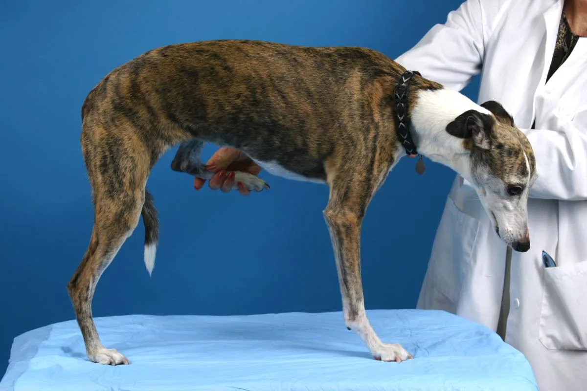
13. Hemistand and Hemiwalk
Evaluate the gait during walking, trotting, and galloping and while turning the patient to the left and right. Hemistanding and hemiwalking (standing and walking on one side) isolates the left and right sides to determine if one side is less coordinated or weaker than the other. Pushing down on the shoulders and hips and observing the resistance to this pressure can also evaluate strength.
Procedure Pearl
Hemistanding and hemiwalking (standing and walking on one side) isolates the left and right sides to determine if one side is less coordinated or weaker than the other.
Spinal Reflexes
The anatomical components of each spinal reflex are specific peripheral sensory nerves, spinal cord segments, motor peripheral nerves, and muscles (indicated in parentheses below). All components must be functional for the spinal reflex to be present. A depressed or absent spinal reflex indicates a lesion in the specific region of the spinal reflex tested. An exaggerated spinal reflex often means a lesion is present somewhere between the brain and the spinal reflex tested.
Thoracic Limb Reflexes
17. Biceps reflex
Place a finger on the biceps tendon and percuss the finger. A brief elbow flexion indicates a normal biceps reflex (C6–C8). The response can be subtle in healthy dogs and cats.
Pelvic Limb Reflexes
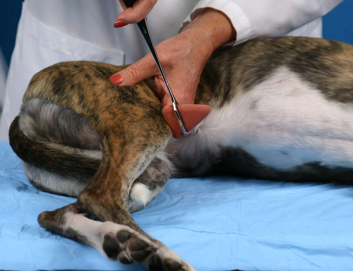
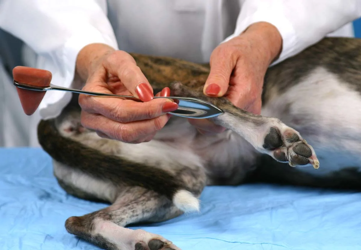
22. Patellar reflex
Percussing the patellar tendon and observing a brief extension of the stifle joint indicates a normal patellar reflex (L4–L5).
Other Examinations
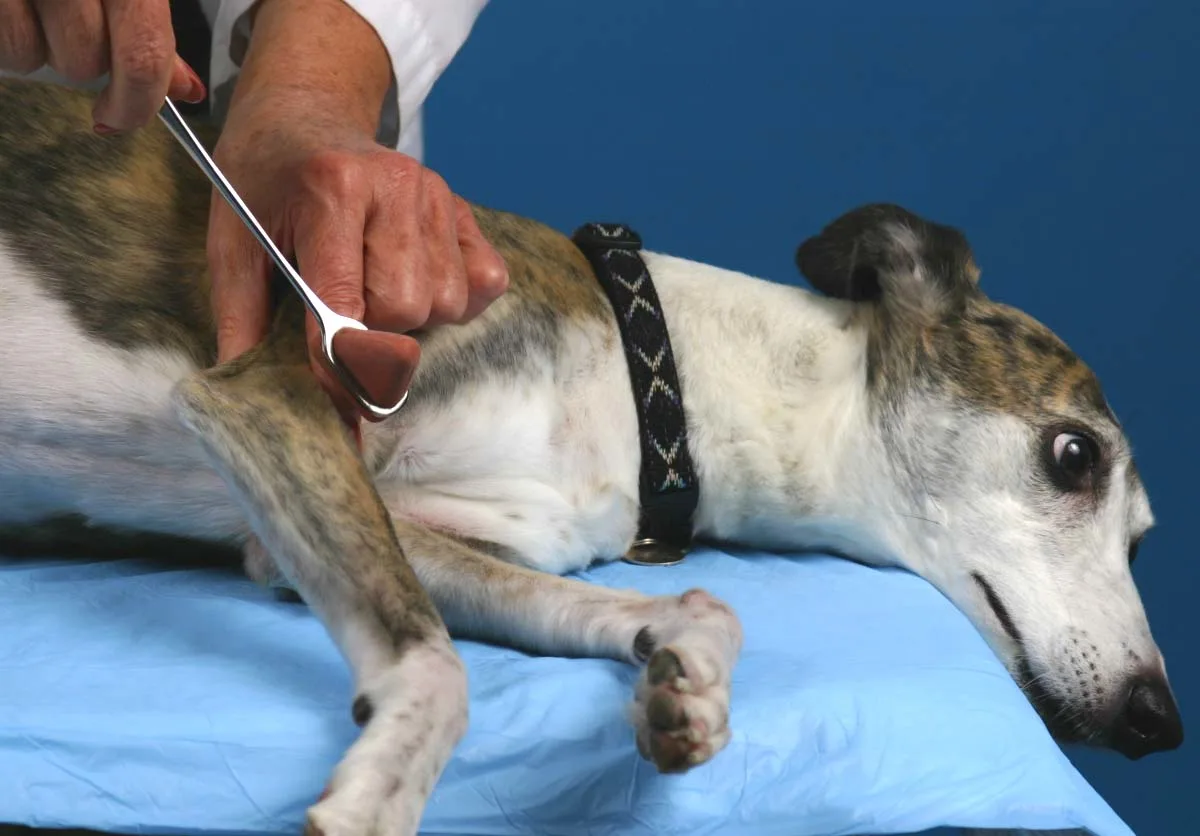
29. Babinski’s sign
Scraping the tip of the percussion hammer proximally on the metacarpal and metatarsal bones elicits slight flexion of the digits. Extension of the digits is a positive Babinski’s sign and indicates a lesion somewhere between the brain and C5 (thoracic limb) or the brain and L5 (pelvic limb).
Author Insight
Extension of the digits is a positive Babinski’s sign and indicates a lesion somewhere between the brain and C5 (thoracic limb) or the brain and L5 (pelvic limb).
Video Series
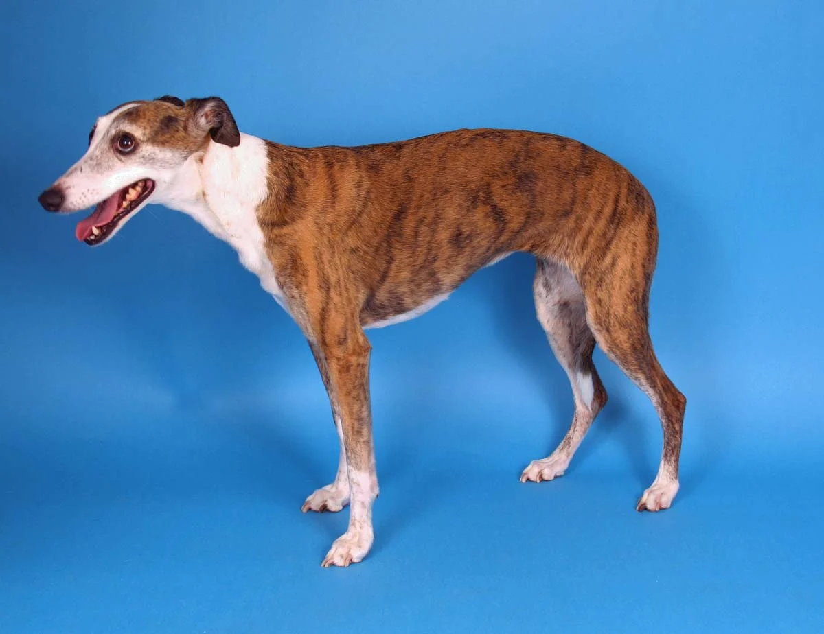
Acknowledgments: In memory of my beloved whippet, “Solo” Windsome’s A Simple Twist of Fate, 1993-2005, the model in the photographs. The author thanks Mark Hoffenberg for the photography.