Refractory Seizures in a Dog

Gracie, a 9-year-old spayed English setter, was referred for refractory epilepsy.
History
Eight months before presentation, Gracie had been diagnosed with probable late-onset cryptogenic epilepsy. However, she had three grand mal seizures within a 24-hour period the week prior to presentation and the day before presentation, two grand mal seizures followed by the onset of status epilepticus in the evening.
Serum phenobarbital levels remained low (<16 µg/mL) despite increasing the dose. The current dose of phenobarbital was 128 mg PO q12h, but the serum drug level was only 10.5 µg/mL. Zonisamide at 100 mg PO q12h was added to help with seizure control.
Examination
On presentation, the dog weighed 45.3 lb and had a rectal temperature of 103.6°F (normal, 100.5°F–102.5°F). No murmur or arrhythmia was noted, and the dog’s respiratory rate and effort were within normal limits.
A firm spherical mass measuring approximately 6 × 5 × 5 cm was palpated in the cranial abdomen and believed to be associated with the left liver lobe. Because seizure activity was refractory to CRI diazepam, CRI propofol was initiated, thereby preventing evaluation of the dog’s full neurologic status.
Diagnostics
An MRI conducted previously did not reveal any intracranial lesions. Serum biochemistry profile results showed hypoglycemia, hypoalbuminemia, hypocalcemia, and decreased concentrations of BUN. The urine protein:creatinine ratio was elevated (See Table 1).
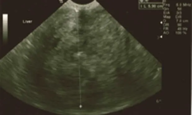
Abdominal ultrasonography revealed a 12-cm encapsulated, hyperechogenic, irregular mass in the cranial abdomen (Figure 1).
The mass was adjacent to the left lateral liver lobe. The remainder of the liver appeared normal, although a minute amount of fluid was present around the mass. The spleen was normal in appearance and the remainder of the abdomen unremarkable.
_
Figure 1. Ultrasound revealed a large, echogenic abdominal mass adjacent to the left lateral liver lobe._
Ask Yourself
What are some differentials for refractory epilepsy in this patient?
What tumors are known to cause hypoglycemia?
Where would these tumors generally be located?
Diagnosis
Leiomyoma & Protein-Losing Nephropathy Likely Secondary to Seizures
Leiomyoma (benign) and leiomyosarcoma (malignant) are composed mainly of smooth muscle (Figure 2) and occur in middle-aged to geriatric dogs with no evident sex predilection. Although there is no definitive breed predilection, poodles and Chihuahuas appear to be overrepresented.1

Figure 2. Leiomyoma has atypical spindloid cells that proliferate as broad interlacing fascicles, mimicking normal smooth muscle. (H&E stain, 40×)
Both tumor types are usually an incidental finding at necropsy. Typical gross appearance of leiomyoma is a semifirm to firm solid mass that is pale pink to tan.1
Certain sites of origin of leiomyosarcomas (eg, duodenum, spleen, liver) appear to have a higher incidence of metastasis in dogs.1 Both leiomyoma and leiomyosarcoma are associated with a paraneoplastic syndrome that causes profound hypoglycemia and concurrent clinical signs. There are several proposed mechanisms of how these tumors create hypoglycemia, but the most likely explanation is production of an insulin-like molecule (eg, insulin-like growth factor 2) that causes altered glucose homeostasis.1-3
GI stromal tumors (GISTs), which cannot be distinguished from smooth muscle tumors with H&E staining, also have reportedly caused hypoglycemia, so additional testing is necessary to confirm smooth muscle differentiation. GISTs are believed to originate from interstitial cells (myofibroblasts), which are precursors to the pacemaker cells of the intestinal wall.1,4 Differentiation is possible with immunohistochemistry. GISTs will likely stain with S100, neuron-specific enolase, synaptophysin, or c-kit protein.1,4 In this case, immunohistochemistry revealed no uptake of c-kit staining, thus supporting the diagnosis of leiomyoma.
GIST = GI stromal tumor, H&E = hematoxylin and eosin
Treatment & Outcome
Leiomyoma is a noninvasive tumor that does not metastasize; therefore, in most cases, surgical resection results in resolution of hypoglycemia. However, in this case the dog was euthanized because ongoing seizures and persistent hypoglycemia despite repeated dextrose administration and CRI diazepam made her a poor surgical candidate.
Definitive diagnosis of leiomyoma was made postmortem, at which time a semifirm 12 × 12 × 8-cm pale pink mass was found wrapped in the root of the mesentery (Figure 3). The only attachment noted was to the blood supply. The liver was diffusely enlarged but normal in appearance.

Figure 3. Gross image of mass, which was dissected, removed from the body, and partially transected. It was semifirm on palpation and pale pink–tan, which is typical of most leiomyomas.
Did You Answer?
Other differentials for refractory epilepsy in this patient include hypoglycemia, hepatic encephalopathy, progression of the previously diagnosed idiopathic epilepsy, brain tumor, inflammatory brain lesion, vascular event, infectious causes, and head trauma.
Types of tumors causing hypoglycemia include leiomyoma, leiomyosarcoma, insulinoma, hepatoma, lymphocytic leukemia, plasma cell myeloma, malignant melanoma, carcinoma, hemangiosarcoma, and malignant lymphoma.3,5
The most commonly reported locations of leiomyoma and leiomyosarcoma are the GI tract, liver, spleen, urinary bladder, and vulva/vagina; however, hypoglycemia has only been reported in tumors occurring in the GI tract, liver, and spleen.
Apparently in dogs, only one other leiomyoma reportedly originated from the vasculature.6
Insulinoma, the most common cause of paraneoplastic hypoglycemia, originates from pancreatic b-islet cells. Malignant melanoma causing hypoglycemia most commonly originates from the oral cavity.7
Hemangiosarcoma is a tumor of vascular origin, and masses are typically found in the liver, spleen, or right atrium. However, hemangiosarcoma has also been reported in the GI tract, CNS, bone, skin, kidney, skeletal muscles, and lung. Malignant lymphoma typically does not form discrete masses and has been reported in almost every body organ.
SARAH E. COCKER, DVM, is an intern at Central Texas Veterinary Specialty Hospital in Austin. Her special interests include hepatobiliary disease, endocrinopathies, and immunemediated disease processes. Dr. Cocker earned her DVM from Oklahoma State University College of Veterinary Medicine and is pursuing a residency in internal medicine.
SHARON K. THEISEN, DVM, DACVIM, practices internal medicine at Central Texas Veterinary Specialty Hospital in Austin, of which she is a founding member. She earned her degree from Texas A&M University College of Veterinary Medicine and completed an internal medicine residency at Ohio State University.