Quiz: Mucous Membrane Evaluation in Dogs
Elizabeth Rozanski, DVM, DACVECC, DACVIM, Cummings School of Veterinary Medicine at Tufts University
Evaluation of mucous membranes—including assessment of color, capillary refill time, and degree of moisture—can help provide vital insights when assessing and diagnosing patients. The author most frequently evaluates the oral mucous membranes, although the conjunctiva and the vulva or prepuce may also be evaluated.
In the author’s experience, mucous membrane color may vary among patients, as some canine breeds such as Weimaraners and red Doberman pinschers have naturally pinker membranes. In addition, some drugs (eg, dexmedetomidine) may alter membrane color.1 Cats have much smaller mouths than do dogs, and interpretation of mucous membrane color may be more challenging than with canine patients.
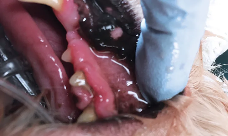
Prompt and markedly delayed capillary refill time are associated with vasodilation (and/or hyperdynamic shock) and vasoconstriction, respectively.2,3 Patients, particularly dogs, may develop vasodilation associated with early shock (eg, sepsis) or heatstroke, which results in mucous membranes appearing brick red in color.3 Conversely, patients in more advanced states of shock will have pale mucous membranes associated with cool extremities that reflect vasoconstriction.3 Critically ill patients may progress from hyperdynamic to hypodynamic shock if illness advances.3 Oral diseases (eg, dental disease) or oral foreign bodies (eg, quills, foxtails) may also result in abnormal oral mucous membrane appearance.3