Physeal Fractures in Small Animals
Mary Sarah Bergh, DVM, MS, DACVS, DACVSMR, Midwest Veterinary Specialists, Milwaukee, Wisconsin

Physeal fractures are common in skeletally immature cats and dogs. Because they can often be difficult to detect on radiographs, a high index of suspicion of physeal injury is warranted in any traumatized puppy or kitten with pain or swelling around the joint. Early identification and timely reduction and stabilization of the fracture, along with careful monitoring, are important for a successful outcome.

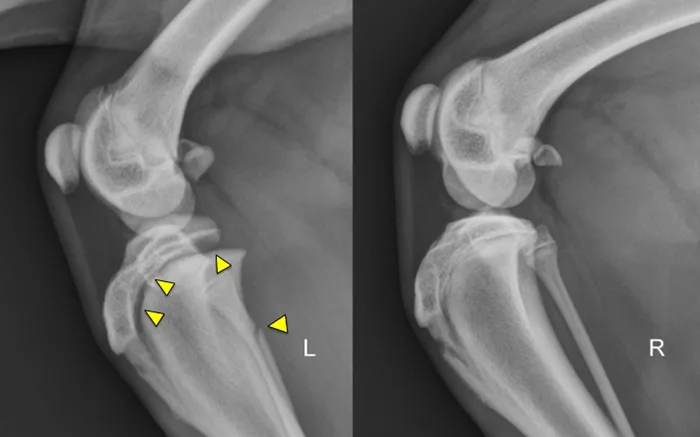
Figure 1 Physeal fractures can occur in any juvenile animal, even with little trauma, because the physis is weaker than the adjacent bone and ligaments. Orthogonal radiographs of any painful joint should be performed. Oblique projections, radiographs of the limb in different positions, and radiographs of the contralateral limb for comparison can all aid in the diagnosis of a physeal fracture. Shown here are lateral stifle radiographs from an 8-month-old mixed breed dog that presented for acute onset of lameness in the left pelvic limb after he slipped earlier that evening; pain was localized to the left stifle. Notice the open physes of the proximal tibia and distal femur, which are normal for a dog of this age. There is physeal separation and cranial displacement of the proximal epiphysis in the left tibia, and a greenstick fracture of the left fibula (arrows).