Image Gallery: Upper Gastrointestinal Endoscopy
Jennifer Gieg, DVM, DACVIM, MedVet, Cincinnati, Ohio
Endoscopy is a valuable tool used to evaluate various upper GI disorders (eg, esophageal strictures, esophagitis, gastric or intestinal ulceration, and inflammatory bowel disease [IBD], neoplasia) as well as for emergency removal of foreign bodies and placement of feeding tubes. Patients with obvious GI signs of distress (eg, vomiting, regurgitation) or more subtle signs (eg, repeated swallowing, inappetence) may benefit from endoscopy.
Pre-Endoscopic Considerations
Pre-endoscopic diagnostic investigation of patients with GI signs should include a CBC, serum chemistry profile, and urinalysis. Other testing to consider before endoscopy may include fecal testing, pancreatic lipase immunoreactivity, vitamin B12 and folate levels, trypsin-like immunoreactivity, adrenocorticotropic hormone stimulation test, and/or serum gastrin levels. Empiric deworming and a novel protein diet or hydrolyzed diet food trial may also be indicated. Diagnostic imaging, including abdominal and thoracic radiography and abdominal ultrasonography may help narrow the list of differentials and indicate whether endoscopy is the best diagnostic tool for the patient. Radiography can help localize disease, while abdominal ultrasonography can help identify lesions and establish those that cannot be treated or are not endoscopically-accessible. Ultrasound-guided fine-needle aspiration of masses or lymph nodes may help identify conditions such as histoplasmosis or lymphoma, and may obviate the diagnostic need for endoscopy or surgery.
Choosing Endoscopy
The decision to pursue endoscopy alone or consider surgical options is dependent on several factors. For example, in patients with an uncomplicated esophageal foreign body (no evidence of perforation) or suspected gastric ulceration, endoscopy may be advisable as a first-line treatment over surgery.1 However, surgery may be indicated in a patient with a distal small intestinal lesion that is inaccessible via endoscopy. For more complicated cases, it is important to identify the location of a lesion in the wall of the target organ and along the length of the GI tract. In patients with marked mural thickening or loss of wall layer, full-thickness surgical biopsy may be preferable over superficial endoscopic biopsy.
Endoscopy is generally safe, with complication rates of less than 1% in humans.2 Foreign body removal carries an increased risk as compared with diagnostic endoscopy; however, the complication rate associated with gastroesophageal foreign body removal is low, with esophageal perforation being the most common complication reported.3 Full upper GI endoscopy should include evaluation of the oral cavity, esophagus, stomach, and small intestine.
Abnormalities & Diagnoses
Endoscopic evaluation of esophageal tissue should note the presence of food and/or fluid, which may suggest reflux, and describe the state of the lower esophageal sphincter (open vs closed). Thorough evaluation of all areas of the stomach includes inspecting the mucosa for abnormalities (eg, punctate areas of hemorrhage, superficial erosion, deep ulceration, erythema, masses). Other abnormalities, including foreign bodies, gastric parasites (eg, Physaloptera spp), or retained gastric contents (eg, food or fluid in a fasted patient), may also be identified. Retroflex examination of the cardia—an area where neoplasia and foreign material may not be easily visualized—is an essential part of complete endoscopy. Clinicians should be mindful of iatrogenic lesions (eg, small hemorrhages from overdistention of the stomach, suction lesions where the scope made contact with mucosa) as well as normal findings that may appear abnormal; for example, mucosa may appear irregular before insufflation of the stomach. Once the scope is beyond the pyloric sphincter, it should be passed as far into the duodenum as the patient and scope size allow.
There is strong evidence of the value of performing both upper GI endoscopy and colonoscopy (the latter with the purpose of obtaining ileal biopsies), even in patients with signs limited to the upper GI tract.4,5 Lack of agreement between duodenal and ileal histopathology in dogs and cats has been reported.4,5 Identifying significant disease is more likely when ileal biopsies are included in the endoscopy plan.
Obtaining tissue every 5 cm from the farthest point of evaluation can provide representative biopsy samples for diagnosis. In dogs, a minimum of 10 to 15 duodenal tissue samples should be collected and at least 6 samples in cats. Six good samples from the stomach are sufficient for either species.6 In most cases, esophageal biopsies should not be attempted. In the case of an esophageal mass, biopsies may be attempted, but caution should be used to avoid perforation. Use of a sharp (disposable or regularly sharpened reusable) biopsy instrument and the largest biopsy unit available can help obtain biopsy samples of adequate quality for diagnosis.
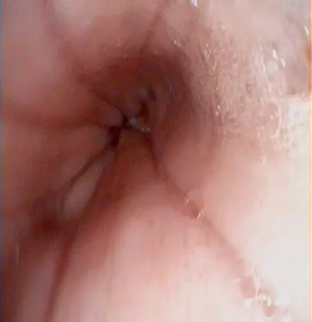
Figure 1A Normal distal esophagus on GI endoscopy