Image Gallery: Corneal Perforations Do Not All Look the Same
Alex Sigmund, DVM, BluePearl Veterinary Partners
Thomas Chen, DVM, DACVO, University of Tennessee
Ocular perforations can occur for a variety of reasons, and their appearance can vary significantly depending on size, etiology, and chronicity.
It is important to recognize when an eye has ruptured or perforated, as surgical correction may be required to maintain the integrity of the eye and ensure the best prognosis for long-term vision and comfort.

Figure 1
Perforation & corneal degenerationred arrows). Calcific degeneration of the cornea is noted in older dogs or results from previous corneal inflammation or injury. As the calcium builds up, it can then slough, resulting in an ulcer. If the ulcer is deep, it could then perforate. A fibrin plug is visible, with pigment likely from secondary iridal prolapse/anterior synechiae (yellow arrow). There is conjunctival hyperemia but no obvious keratitis or neovascularization; in addition, except for the degenerative change, the cornea is largely clear. The degenerative changes to the cornea are chronic and progressive, but the perforation is acute.
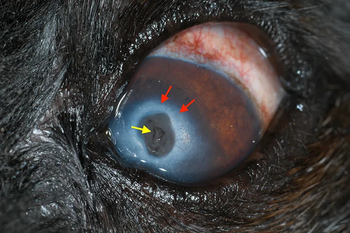
Corneal perforation in a dog with corneal degeneration, most likely calcific degeneration (red arrows). Calcific degeneration of the cornea is noted in older dogs or results from previous corneal inflammation or injury. As the calcium builds up, it can then slough, resulting in an ulcer. If the ulcer is deep, it could then perforate. A fibrin plug is visible, with pigment likely from secondary iridal prolapse/anterior synechiae (yellow arrow). There is conjunctival hyperemia but no obvious keratitis or neovascularization; in addition, except for the degenerative change, the cornea is largely clear. The degenerative changes to the cornea are chronic and progressive, but the perforation is acute.
Images with permission, courtesy University of Tennessee College of Veterinary Medicine